

Quadriceps Performance and Running Biomechanics Influence Femur BMD Changes after ACL Reconstruction in Collegiate Athletes
- PMID: 37101347
- PMCID: PMC10523868
- DOI: 10.1249/MSS.0000000000003186
Quadriceps Performance and Running Biomechanics Influence Femur BMD Changes after ACL Reconstruction in Collegiate Athletes
Abstract
Purpose: Reduced bone mineral density of the distal femur (BMD DF ) can persist long term after anterior cruciate ligament reconstruction (ACLR), even in athletes who return to high levels of competition. These deficits may have implications for the onset and progression of knee osteoarthritis. It is unknown if clinically modifiable factors are associated with losses in BMD DF . This study evaluated the potential influence of knee extensor peak torque (PT), rate of torque development (RTD), as well as peak knee flexion (PKF) angle and peak knee extensor moment (PKEM) during running, on longitudinal changes in BMD DF post-ACLR.
Methods: After ACLR, 57 Division I collegiate athletes underwent serial whole-body dual-energy x-ray absorptiometry (DXA) scans between 3 and 24 months post-ACLR. Of these, 43 athletes also had isometric knee extensor testing (21 female, 105 observations), and 54 had running analyses (26 female, 141 observations). Linear mixed-effects models, controlling for sex, assessed the influence of surgical limb quadriceps performance (PT and RTD), running mechanics (PKF and PKEM), and time post-ACLR on BMD DF (5% and 15% of femur length). Simple slope analyses were used to explore interactions.
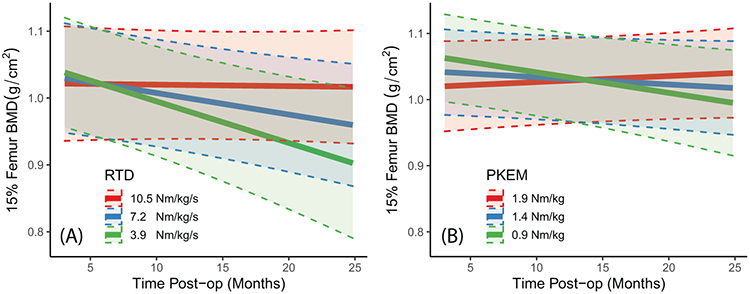
Results: Athletes with RTD less than 7.20 (N·m)·kg -1 ·s -1 (mean) at 9.3 months post-ACLR demonstrated significant decreases in 15% BMD DF over time ( P = 0.03). Athletes with PKEM during running less than 0.92 (N·m)·kg -1 (-1 SD below mean) at 9.8 months post-ACLR demonstrated significant decreases in 15% BMD DF over time ( P = 0.02). Significant slopes were not detected at -1 SD below the mean for PT (1.75 (N·m)·kg -1 , P = 0.07) and PKF (31.3°, P = 0.08).
Conclusions: Worse quadriceps RTD and running PKEM were associated with a greater loss of BMD DF between 3 and 24 months post-ACLR.
Copyright © 2023 by the American College of Sports Medicine.
Conflict of interest statement
Research reported in the publication was supported by the National Institute of Health through award number TL1TR002375 and T32AG000213, as well as the University of Wisconsin-Madison Department of Orthopedics and Rehabilitation Research Grant. The content is solely the responsibility of the authors and does not necessarily represent the official views of the NIH.
Dr. Heiderscheit declares a potential conflict of interest not directly related to this work (ownership interest in Science of Running Medicine, LLC, advisory board member for Springbok Analytics, consultant for Biocore, Inc., and research support (to institution) from National Football League). Dr. Binkley declares a potential conflict of interest not directly related to this work (Consultant to Amgen and Research Support (to Institution): Radius. Dr. Scerpella declares a potential conflict of interest not directly related to this work (Johnson and Johnson Medical Device Business Services - Development of Educational, Marketing Collateral/Training Materials (Limited IP)). The results of the study are presented clearly, honestly, and without fabrication, falsification, or inappropriate data manipulation. The results of the present study do not constitute endorsement by the American College of Sports Medicine.
Figures


References
-
- Kannus P, Sievänen H, Järvinen M, Heinonen A, Oja P, Vuori I. A cruciate ligament injury produces considerable, permanent osteoporosis in the affected knee. J Bone Miner Res. 1992;7(12):1429–34. - PubMed
-
- Nyland J, Fisher B, Brand E, Krupp R, Caborn DNM. Osseous deficits after anterior cruciate ligament injury and reconstruction: a systematic literature review with suggestions to improve osseous homeostasis. Arthroscopy. 2010;26(9):1248–57. - PubMed
-
- Mündermann A, Payer N, Felmet G, Riehle H. Comparison of volumetric bone mineral density in the operated and contralateral knee after anterior cruciate ligament and reconstruction: a 1-year follow-up study using peripheral quantitative computed tomography. J Orthop Res. 2015;33(12):1804–10. - PubMed
-
- Rittweger J, Reeves ND, Narici MV, Belavý DL, Maganaris CN, Maffulli N. Persisting side-to-side differences in bone mineral content, but not in muscle strength and tendon stiffness after anterior cruciate ligament reconstruction. Clin Physiol Funct Imaging. 2011;31(1):73–9. - PubMed
-
- van Meer BL, Waarsing JH, van Eijsden WA, et al. Bone mineral density changes in the knee following anterior cruciate ligament rupture. Osteoarthritis Cartilage. 2014;22(1):154–61. - PubMed
Publication types
MeSH terms
Grants and funding
LinkOut - more resources
Full Text Sources
Miscellaneous
