. 2023 Feb 15;3(2):158-173.
doi: 10.1021/acsbiomedchemau.2c00067. eCollection 2023 Apr 19.

Controlling Amyloid Beta Peptide Aggregation and Toxicity by Protease-Stable Ligands

Affiliations

Controlling Amyloid Beta Peptide Aggregation and Toxicity by Protease-Stable Ligands

Rathnam Mallesh et al. ACS Bio Med Chem Au. .

Abstract

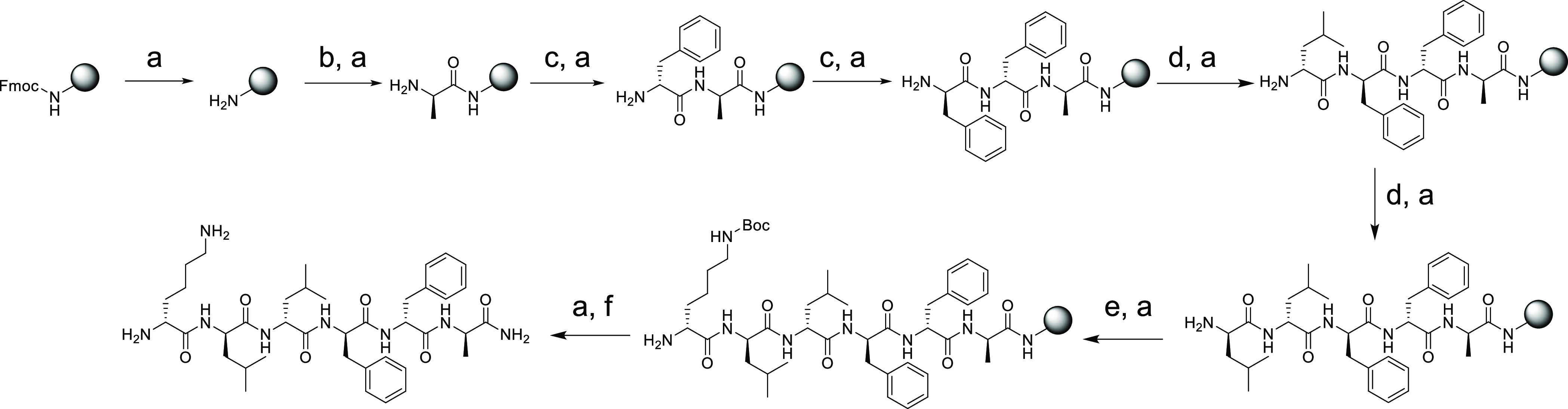
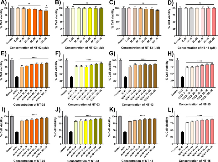
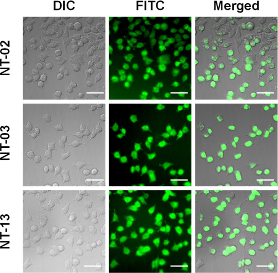
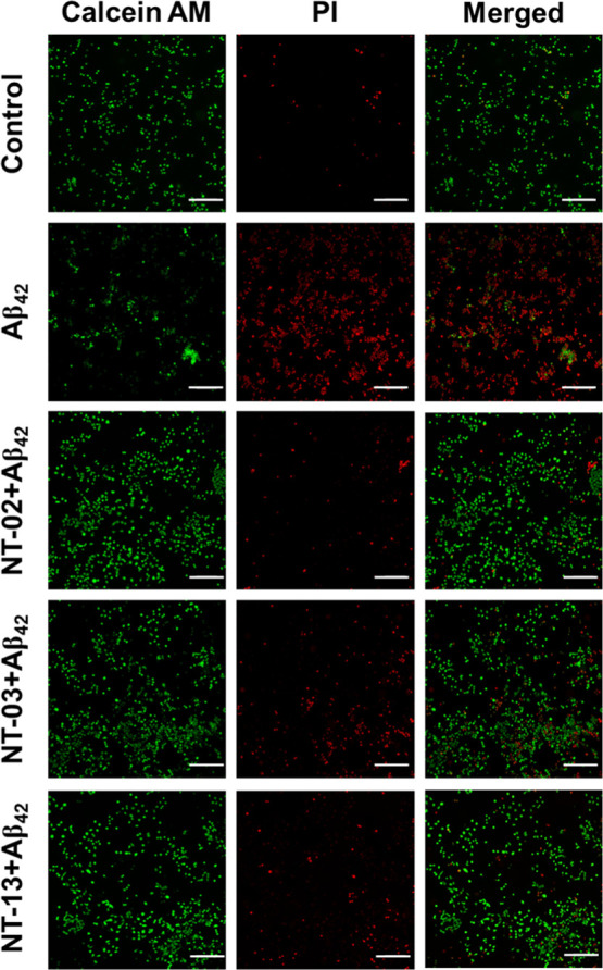
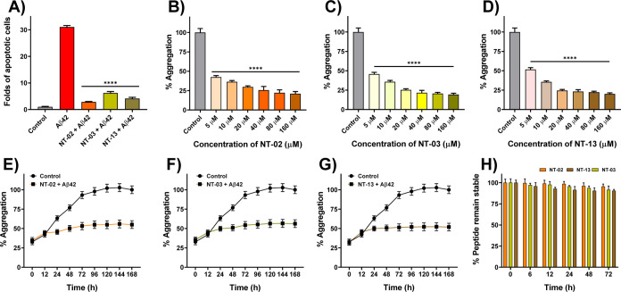
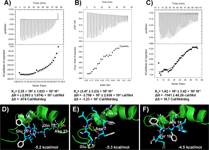
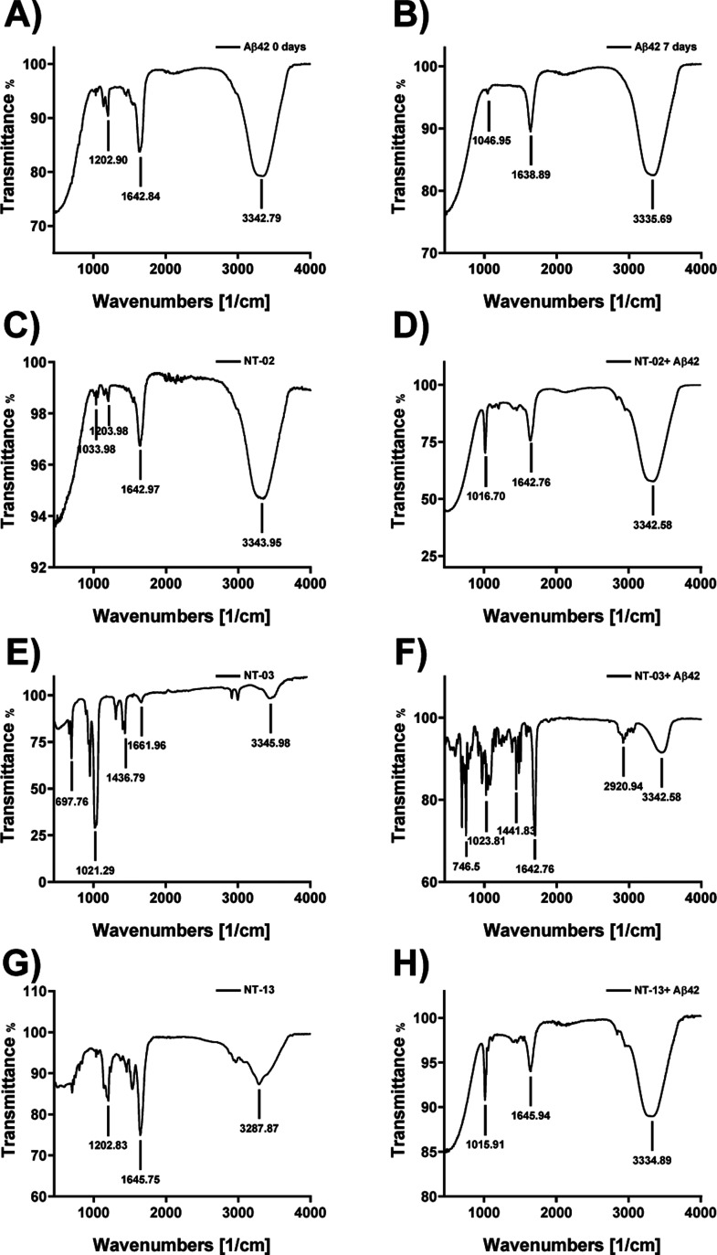
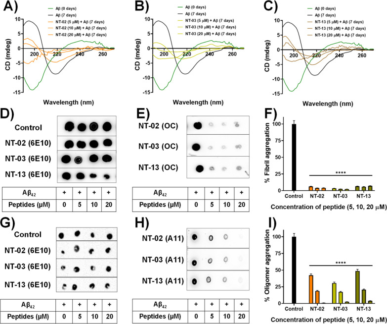
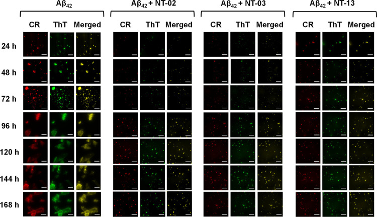
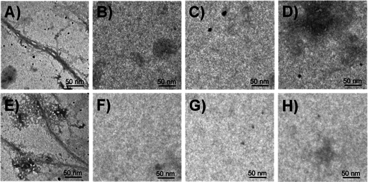
Polymerization of soluble amyloid beta (Aβ) peptide into protease-stable insoluble fibrillary aggregates is a critical step in the pathogenesis of Alzheimer's disease (AD). The N-terminal (NT) hydrophobic central domain fragment 16KLVFF20 plays an important role in the formation and stabilization of β-sheets by self-recognition of the parent Aβ peptide, followed by aggregation of Aβ in the AD brain. Here, we analyze the effect of the NT region inducing β-sheet formation in the Aβ peptide by a single amino acid mutation in the native Aβ peptide fragment. We designed 14 hydrophobic peptides (NT-01 to NT-14) by a single mutation at 18Val by using hydrophobic residues leucine and proline in the natural Aβ peptide fragment (KLVFFAE) and analyzed its effect on the formation of Aβ aggregates. Among all these peptides, NT-02, NT-03, and NT-13 significantly affected the Aβ aggregate formation. When the NT peptides were coincubated with the Aβ peptide, a significant reduction in β-sheet formation and increment in random coil content of Aβ was seen, confirmed by circular dichroism spectroscopy and Fourier transform infrared spectroscopy, followed by the reduction of fibril formation measured by the thioflavin-T (ThT) binding assay. The aggregation inhibition was monitored by Congo red and ThT staining and electron microscopic examination. Moreover, the NT peptides protect the PC-12 differentiated neurons from Aβ-induced toxicity and apoptosis in vitro. Thus, manipulation of the Aβ secondary structure with protease-stable ligands that promote the random coil conformation may provide a tool to control the Aβ aggregates observed in AD patients.


Conflict of interest statement

The authors declare no competing financial interest.

Figures

Scheme 1
Scheme 1. Synthetic Scheme for the Peptides by Using Rink Amide Resin Exemplified by Peptide NT-02
Reagents and conditions: (a) 20% piperidine in DMF, 3 min (two times); (b) Fmoc-d-Ala-OH (10 equiv), HBTU (10 equiv), DIPEA (10 equiv), DMF, 7 min; (c) Fmoc-d-Phe-OH (5 equiv), HBTU (5 equiv), DIPEA (10 equiv), DMF, 7 min; (d) Fmoc-d-Leu-OH (5 equiv), HBTU (5 equiv), DIPEA (10 equiv), DMF, 7 min; (e) Fmoc-d-Lys (Boc)-OH (5 equiv), HBTU (5 equiv), DIPEA (10 equiv), DMF, 7 min; (f) 92.5% TFA, 2.5% water, 2.5% phenol, 2.5% 1.2-ethylenedithiol, 12 h.
Figure 1
Figure 1
Cellular viability of peptides (A) NT-02, (B) NT-03, (C) NT-13, and (D) positive control NT-15 (KLVFF) in PC-12-derived neurons by using the MTT assay. Effects of NT peptides on the Aβ42 peptide-induced cytotoxicity in PC-12-derived neurons. (E) PC-12-derived neurons were treated with the Aβ42 peptide (5 μM) alone and Aβ42 peptide with (E) NT-02, (F) NT-03, (G) NT-13 peptide, and (H) positive control NT-15 for 24 h after which their ability to reduce MTT was measured. Cellular viability of PC12-derived neurons after being treated with Aβ42 aggregates (5 μM) alone and Aβ42 aggregates with (I) NT-02, (J) NT-03, (K) NT-13 peptides, and (L) positive control NT-15 for 24 h after which their ability to reduce MTT was measured. Error bars represent mean ± standard deviation (SD), n = 3. Statistical data were analyzed by a one-way ANOVA test by the multiple comparison tests (*p < 0.05, **p < 0.01, ***p < 0.001, ****p < 0.0001, vs Aβ42) using software (GraphPad Prism, ISI, San Diego, CA).
Figure 2
Figure 2
Cellular uptake images of 5(6)-carboxyfluorescein-attached NT-02, NT-03, and NT-13 peptides in differentiated PC-12-derived neurons reveal a significant cellular uptake. The scale bar corresponds to 50 μm.
Figure 3
Figure 3
Cell apoptosis. Fluorescence images of differentiated PC-12-derived neuronal cells stained with Calcein AM and PI for normal and late apoptotic cell visualization. Initiation of apoptosis by Aβ42 peptide treatment compared to the control and rescue by NT-02, NT-03, and NT-13 peptides. The scale bar corresponds to 200 μm.
Figure 4
Figure 4
(A) Quantification of percentage apoptotic cells of different treatments shows the significant rescue of cells from Aβ42-induced apoptosis by NT-02, NT-03, and NT-13 peptides. Effects of NT peptides on Aβ42 aggregation measured by the ThT fluorescence assay. (B) Peptides NT-02, (C) NT-03, and (D) NT-13 in Aβ42 aggregation. The control sample represents the Aβ42 peptide (5 μM) alone. (E–G) Effects of peptides NT-02, NT-03, and NT-13 on Aβ42 aggregation for 7 days. Aβ42 (5 μM) and peptide at a 10 μM concentration. Control sample represents the Aβ42 peptide (5 μM) alone. (H) Serum stability of NT peptides in human serum up to 24 h. Error bars represent mean ± standard deviation (SD), n = 3. Statistical data were analyzed by a one-way ANOVA test by the multiple comparison tests (*p < 0.05, **p < 0.01, ***p < 0.001, ****p < 0.0001, vs Aβ42) using software GraphPad Prism (ISI, San Diego, CA).
Figure 5
Figure 5
ITC experiment of NT peptides showing interaction with the Aβ42 peptide. (A) NT-02 peptide shows an affinity of 2.25 × 10⁶ ± 1.023 × 10⁵ M–1, (B) NT-03 peptide shows an affinity of (3.47 ± 1.23) × 106 M–1, and (C) NT-13 peptide shows an affinity of 1.42 × 10⁵ ± 3.42 × 10⁴ M–1. Molecular docking images of NT peptides with the Aβ42 peptide (PDB ID: 1Z0Q). (D) Docking studies of the NT-02 peptide with the Aβ42 peptide show H-bonding interaction with a −5.2 kcal/mol binding energy. (E) Docking studies of NT-03 with Aβ42 and showing H-bonding interactions with −5.3 kcal/mol binding energy. (F) Docking studies of NT-13 with Aβ42 and showing H-bonding interaction with a −4.5 kcal/mol binding energy.
Figure 6
Figure 6
Analysis of β-sheet content by FTIR spectroscopy. FTIR spectrum of the (A) Aβ42 peptide alone for 0 day incubation. The spectrum shows the Aβ42 peptide in α-helix conformation. (B) Aβ42 peptide alone upon 7 days of incubation. The spectrum shows that the Aβ42 peptide converted into β-sheet conformation. (C,E,G) FTIR spectrum of peptide NT-02, 03, and 13 alone for 7 days of incubation. The spectrum shows that NT peptides in α-helix conformation. (D,F,H) FTIR spectrum of Aβ42 with NT-02, 03, and 13 for 7 days of incubation. The spectrum shows the Aβ42 peptide in α-helix conformation.
Figure 7
Figure 7
CD spectroscopic studies showing β-sheet content and the inhibitory effects of peptides (A) NT-02, (B) NT-03, and (C) NT-13. CD spectra show the Aβ42 sample at t = 0 h (green), and the Aβ42 sample at t = 7 days (black). Dot blot assay for monitoring Aβ42 aggregation. (D) Dot blot image for Aβ42 content detected by 6E10. (E) Dot blot image for Aβ42 fibrillary aggregation inhibition in the presence of NT peptides detected by OC. (F) Bar diagram of Aβ42 fibrillary aggregation inhibition by NT peptides. (G) Dot blot image for Aβ42 content detected by 6E10. (H) Dot blot image for Aβ42 oligomeric aggregation inhibition in the presence of NT peptides detected by A11. (I) Bar diagram of Aβ42 oligomer inhibition by NT peptides. Error bars represent mean ± standard deviation (SD), n = 3. Statistical data were analyzed by a one-way ANOVA test by the multiple comparison test (*p < 0.05, **p < 0.01, ***p < 0.001, ****p < 0.0001, vs control) using software (GraphPad Prism, ISI, San Diego, CA).
Figure 8
Figure 8
CR and ThT binding to Aβ42 aggregates. Fluorescence microscopical images show the presence of Aβ42 aggregates in all samples. Fluorescence microscopic images of 10 μM Aβ42 peptide in the presence of 1 μM CR (column 1) and ThT (column 2). Images clearly indicate that NT peptides inhibit the formation of Aβ42 aggregates. The scale bar corresponds to 200 μm.
Figure 9
Figure 9
HR-TEM images. (A,E) Aβ42 peptide fibrils after 7 days of incubation of Aβ42 alone at 37 °C. Absence of the fibril structure after coincubation of the Aβ42 peptide with (B) NT-02, (C) NT-03, and (D) NT-13 peptides at 37 °C for 7 days. Peptides (F) NT-02, (G) NT-03, and (H) NT-13 incubated alone for 7 days, and no fibril formation was observed.

Similar articles

Cited by

References

    1. Wong C. W.; Quaranta V.; Glenner G. G. Neuritic Plaques and Cerebrovascular Amyloid in Alzheimer Disease Are Antigenically Related. Proc. Natl. Acad. Sci. 1985, 82, 8729–8732. 10.1073/pnas.82.24.8729. - DOI - PMC - PubMed
    1. Glenner G. G.; Wong C. W. Alzheimer’s Disease: Initial Report of the Purification and Characterization of a Novel Cerebrovascular Amyloid Protein. Biochem. Biophys. Res. Commun. 1984, 120, 885–890. 10.1016/s0006-291x(84)80190-4. - DOI - PubMed
    1. Roher A. E.; Lowenson J. D.; Clarke S.; Woods A. S.; Cotter R. J.; Gowing E.; Ball M. J. Beta-Amyloid-(1-42) Is a Major Component of Cerebrovascular Amyloid Deposits: Implications for the Pathology of Alzheimer Disease. Proc. Natl. Acad. Sci. 1993, 90, 10836–10840. 10.1073/pnas.90.22.10836. - DOI - PMC - PubMed
    1. Selkoe D. J. Amyloid β-Protein and the Genetics of Alzheimer’s Disease. J. Biol. Chem. 1996, 271, 18295–18298. 10.1074/jbc.271.31.18295. - DOI - PubMed
    1. Terzi E.; Hölzemann G.; Seelig J. Self-Association of β-Amyloid Peptide (1–40) in Solution and Binding to Lipid Membranes. J. Mol. Biol. 1995, 252, 633–642. 10.1006/jmbi.1995.0525. - DOI - PubMed