

Acute Peritonitis Is Not Always a Surgical Fix: A Rare Case of Mixed Connective Tissue Disease Presenting as Polyserositis
- PMID: 37102006
- PMCID: PMC10123235
- DOI: 10.7759/cureus.36652
Acute Peritonitis Is Not Always a Surgical Fix: A Rare Case of Mixed Connective Tissue Disease Presenting as Polyserositis
Abstract
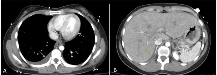
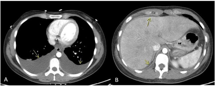
Mixed connective tissue disease (MCTD) is a complex rheumatologic condition whose diagnosis often presents a challenge to even specialists in the field. Many cases are therefore underrecognized or misdiagnosed due to the heterogeneity of the presentation and manifestations. This report highlights the intricacies of diagnosing a case of MCTD when the presenting symptom is atypical. Herein, we present a case of a young girl who had severe abdominal pain, initially concerning for acute peritonitis from cholecystitis, and was found to have polyserositis affecting the pleural space, pericardium, peritoneum and pelvis secondary to mixed connective tissue disease and adrenal insufficiency.
Keywords: acute abdomen; autoimmune disease; mixed-connective-tissue-disease; peritonitis; polyserositis; rheumatological disease; serositis.
Copyright © 2023, Tagliaferri et al.
Conflict of interest statement
The authors have declared that no competing interests exist.
Figures
Similar articles
-
Seropositive Neuromyelitis Optica in a Case of Undiagnosed Ankylosing Spondylitis: A Neuro-Rheumatological Conundrum.Qatar Med J. 2022 Jul 7;2022(3):29. doi: 10.5339/qmj.2022.29. eCollection 2022. Qatar Med J. 2022. PMID: 35864917 Free PMC article.
-
Polyserositis and Acute Acalculous Cholecystitis: An Uncommon Manifestation of Undiagnosed Systemic Lupus Erythematosus.Cureus. 2019 Jun 14;11(6):e4899. doi: 10.7759/cureus.4899. Cureus. 2019. PMID: 31423378 Free PMC article.
-
Anakinra effectiveness in refractory polyserositis: An Italian multicenter study.Joint Bone Spine. 2022 Mar;89(2):105299. doi: 10.1016/j.jbspin.2021.105299. Epub 2021 Oct 14. Joint Bone Spine. 2022. PMID: 34656754
-
Life-threatening acute pneumonitis in mixed connective tissue disease: a case report and literature review.Wien Klin Wochenschr. 2015 Oct;127(19-20):792-4. doi: 10.1007/s00508-015-0823-6. Epub 2015 Jul 4. Wien Klin Wochenschr. 2015. PMID: 26142172 Review.
-
Serositis: comparative analysis of histological findings and pathogenetic mechanisms in nonbacterial serosal inflammation.Perit Dial Int. 1993;13(4):256-69. Perit Dial Int. 1993. PMID: 8241326 Review.
References
-
- Mixed connective tissue disease. Venables PJ. Lupus. 2006;15:132–137. - PubMed
Publication types
LinkOut - more resources
Full Text Sources