. 2023 Apr;11(4):e832.
doi: 10.1002/iid3.832.

Citrulline inhibits LPS-induced pyroptosis of RAW264.7 macrophages through NF-κB signaling pathway

Affiliations

Citrulline inhibits LPS-induced pyroptosis of RAW264.7 macrophages through NF-κB signaling pathway

Li Yin et al. Immun Inflamm Dis. 2023 Apr.

Abstract

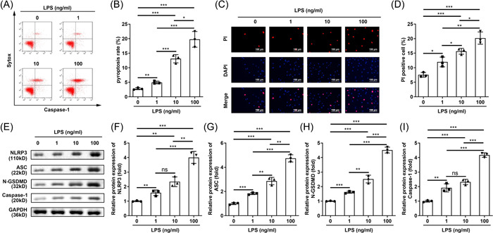
Background: The aim of this study was to investigate the effect of citrulline on the pyroptosis of mouse macrophage RAW264.7 and the mechanism. We investigated the effect of citrulline on pyroptosis of RAW264.7 cell induced by lipopolysaccharide (LPS), and the modulation of nuclear factor-kappaB (NF-κB) signaling.

Methods: Pyroptosis was evaluated using flow cytometry and caspase-1/sytox double staining. Cell counting kit-8 assay was performed to evaluate cell viability.

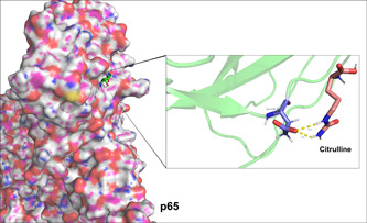
Results: Citrulline promoted cell viability and inhibited the pyroptosis of RAW264.7 cell stimulated by LPS. Furthermore, citrulline inactivated NF-κb/p65 signaling pathway by suppressing p65 nuclear translocation induced by LPS. An NF-κb signaling pathway activator, betulinic acid, reversed the inhibition of pyroptosis induced by citrulline.

Conclusion: Citrulline inhibited LPS-induced pyrophosis, which may be closely related to the inactivation of NF-κB/p65 signaling pathway.

Keywords: NF-κB; citrulline; macrophage; p65; pyroptosis.


Conflict of interest statement

The authors declare no conflict of interest.

Figures

Figure 1
Figure 1
LPS induces pyroptosis of RAW264.7 macrophages. (A, B) Caspase‐1/sytox green staining and flow cytometry was used to detect the pyroptosis of LPS‐treated RAW264.7 cells. (C, D) DAPI/PI double staining assay was performed to detect cell death (magnification: ×100). (E) Western blot analysis was used to evaluate the expression of NLRP3, ASC, N‐GSDMD, and caspase‐1 in LPS‐treated RAW264.7 cells. N = 3. Data were analyzed using one‐way ANOVA followed by Tukey's test Protein levels of (F) NLRP3, (G) ASC, (H) N‐GSDMD, and (I) caspase‐1 were quantified. *p < .05, **p < .01, ***p < .001. ANOVA, analysis of variance; LPS, lipopolysaccharide.
Figure 2
Figure 2
Citrulline inhibits pyroptosis of RAW264.7 macrophages stimulated by LPS. Molecular structure of citrulline. (B) Cell viability of RAW264.7 cells treated with LPS and citrulline was measured by CCK‐8 assay. (C, D) Cell pyroptosis of RAW264.7 cells treated with LPS and citrulline was evaluated by flow cytometry. (E, F) Cell death of RAW264.7 cells treated with LPS and citrulline was evaluated by DAPI/PI double staining assay (magnification: ×100). (G–K) NLRP3, ASC, N‐GSDMD, and caspase‐1 protein levels in RAW264.7 cells treated with LPS and citrulline were measured by western blot. N = 3. Data were analyzed using one‐way ANOVA followed by Tukey's test. **p < .01, ***p < .001. ANOVA, analysis of variance; LPS, lipopolysaccharide, ns, no statistical.
Figure 3
Figure 3
Molecular docking between citrulline and p65.
Figure 4
Figure 4
Citrulline suppresses NF‐κB/p65 nuclear translocation in RAW264.7 macrophages induced by LPS. (A–D) The levels of IκB‐α and p65 and their phosphorylated form proteins were quantified by western blot in RAW264.7 cells treated with LPS and citrulline. (E) The protein level of p65 in RAW264.7 cells treated with LPS and citrulline was assessed by immunofluorescence staining (magnification: ×400). N = 3. Data were analyzed using one‐way ANOVA followed by Tukey's test. **p < .01, ***p < .001. ANOVA, analysis of variance; LPS, lipopolysaccharide; NF‐κB, nuclear factor‐kappaB.
Figure 5
Figure 5
Citrulline suppresses pyroptosis of RAW264.7 macrophages stimulated by LPS by inactivating NF‐κB/p65 pathway. (A) Cell viability of RAW264.7 cells treated with LPS, citrulline, and BA was measured by CCK‐8 assay. (B, C) Cell pyroptosis of RAW264.7 cells treated with LPS, citrulline, and BA was evaluated by flow cytometry. (D, E) Cell death of RAW264.7 cells treated with LPS, citrulline, and BA was evaluated by DAPI/PI double staining assay (magnification: ×100). (F–J) NLRP3, ASC, N‐GSDMD, and caspase‐1 protein levels in RAW264.7 cells treated with LPS, citrulline, and BA were measured by western blot. N = 3. Data were analyzed using one‐way ANOVA followed by Tukey's test. **p < .01, ***p < .001. ANOVA, analysis of variance; BA, betulinic acid; LPS, lipopolysaccharide; NF‐κB, nuclear factor‐kappaB.

References

    1. Li L, Wan G, Han B, Zhang Z. Echinacoside alleviated LPS‐induced cell apoptosis and inflammation in rat intestine epithelial cells by inhibiting the mTOR/STAT3 pathway. Biomed Pharmacother. 2018;104:622‐628. - PubMed
    1. Fritsche KL. The science of fatty acids and inflammation. Adv Nutr. 2015;6(3):293S‐301S. - PMC - PubMed
    1. Arulselvan P, Fard MT, Tan WS, et al. Role of antioxidants and natural products in inflammation. Oxid Med Cell Longevity. 2016;2016:5276130. - PMC - PubMed
    1. Zhong J, Shi G. Editorial: regulation of inflammation in chronic disease. Front Immunol. 2019;10:737. - PMC - PubMed
    1. Kesavardhana S, Malireddi R, Kanneganti TD. Caspases in cell death, inflammation, and pyroptosis. Annu Rev Immunol. 2020;38:567‐595. - PMC - PubMed

Publication types