. 2023 Jun 15;11(3):e0424222.
doi: 10.1128/spectrum.04242-22. Epub 2023 Apr 27.

Hyphal Growth in Trichosporon asahii Is Accelerated by the Addition of Magnesium

Affiliations

Hyphal Growth in Trichosporon asahii Is Accelerated by the Addition of Magnesium

Keita Aoki et al. Microbiol Spectr. .

Abstract

Fungal dimorphism involves two morphologies: a unicellular yeast cell and a multicellular hyphal form. Invasion of hyphae into human cells causes severe opportunistic infections. The transition between yeast and hyphal forms is associated with the virulence of fungi; however, the mechanism is poorly understood. Therefore, we aimed to identify factors that induce hyphal growth of Trichosporon asahii, a dimorphic basidiomycete that causes trichosporonosis. T. asahii showed poor growth and formed small cells containing large lipid droplets and fragmented mitochondria when cultivated for 16 h in a nutrient-deficient liquid medium. However, these phenotypes were suppressed via the addition of yeast nitrogen base. When T. asahii cells were cultivated in the presence of different compounds present in the yeast nitrogen base, we found that magnesium sulfate was a key factor for inducing cell elongation, and its addition dramatically restored hyphal growth in T. asahii. In T. asahii hyphae, vacuoles were enlarged, the size of lipid droplets was decreased, and mitochondria were distributed throughout the cell cytoplasm and adjacent to the cell walls. Additionally, hyphal growth was disrupted due to treatment with an actin inhibitor. The actin inhibitor latrunculin A disrupted the mitochondrial distribution even in hyphal cells. Furthermore, magnesium sulfate treatment accelerated hyphal growth in T. asahii for 72 h when the cells were cultivated in a nutrient-deficient liquid medium. Collectively, our results suggest that an increase in magnesium levels triggers the transition from the yeast to hyphal form in T. asahii. These findings will support studies on the pathogenesis of fungi and aid in developing treatments. IMPORTANCE Understanding the mechanism underlying fungal dimorphism is crucial to discern its invasion into human cells. Invasion is caused by the hyphal form rather than the yeast form; therefore, it is important to understand the mechanism of transition from the yeast to hyphal form. To study the transition mechanism, we utilized Trichosporon asahii, a dimorphic basidiomycete that causes severe trichosporonosis since there are fewer studies on T. asahii than on ascomycetes. This study suggests that an increase in Mg2+, the most abundant mineral in living cells, triggers growth of filamentous hyphae and increases the distribution of mitochondria throughout the cell cytoplasm and adjacent to the cell walls in T. asahii. Understanding the mechanism of hyphal growth triggered by Mg2+ increase will provide a model system to explore fungal pathogenicity in the future.

Keywords: Trichosporon asahii; fungal dimorphism; hyphal growth; magnesium.


Conflict of interest statement

The authors declare no conflict of interest.

Figures

FIG 1
FIG 1
T. asahii formed small cells containing large lipid droplets and showed slow growth in Sabouraud medium. (A) Lipid droplets and DNA of JCM 2466 cells cultivated in YPD and Sabouraud media for 16 h at 25°C were stained with Nile red and DAPI, respectively. NS, not-stained control. (B) The cell surrounded by a dotted line in panel A is enlarged. Lipid droplets, which are not stained by DAPI, are indicated by arrows. Scale bar is 10 μm. (C) Growth rates of JCM 2466 cells cultivated in three liquid media, YPD medium (blue line), Sabouraud medium (green line), and Sabouraud medium containing YNB (orange line), were measured every 10 min using a bio-photorecorder to determine the OD660 for 72 h at 25°C. DIC, differential interference contrast microscopy; YPD, yeast extract-peptone-dextrose medium; YNB, yeast nitrogen base; OD660, optical density at 660 nm.
FIG 2
FIG 2
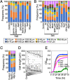
Magnesium sulfate treatment induced hyphal growth in T. asahii. (A) The length of JCM 2466 cells was measured after cultivation in the control, Sabouraud medium containing each ingredient (amino acids, trace elements, salts, or vitamins) of YNB, and Sabouraud medium containing each dropout of the ingredient and then shown as the cell length classification. The amino acids were l-histidine monohydrochloride, ld-methionine, and ld-tryptophan. The trace elements were boric acid, manganese sulfate, zinc sulfate, ferric chloride, sodium molybdate, potassium iodide, and copper sulfate. The salts were ammonium sulfate, monopotassium phosphate, magnesium sulfate, sodium chloride, and calcium chloride. The vitamins were inositol, niacin, pyridoxine, thiamine, calcium pantothenate, riboflavin, p-aminobenzoic acid, folic acid, and biotin. (B) Lengths of JCM 2466 cells were measured after cultivation in control, Sabouraud medium containing each ingredient [KH2PO4, CaCl2, NaCl, (NH4)2SO4, or MgSO4] of the salt group, and Sabouraud medium containing a dropout of each ingredient of the salt group and then shown as the cell length classification. Δ indicates dropout of the corresponding ingredient. (C) Lengths of JCM 2466 cells cultivated in the control medium, Sabouraud medium containing 4.15 mM MgSO4, and Sabouraud medium containing MgCl2 were measured and shown as the cell length classification. (D) Lengths of JCM 2466 cells were measured after cultivation in Sabouraud medium containing MgSO4 at different concentrations for 16 h at 25°C. The concentration of MgSO4 in the ×1 column was 4.15 mM. The dilution series of MgSO4 was 1/10 (415 nM), 1/100 (41.5 nM), 1/200 (20.75 nM), 1/400 (10.38 nM), 1/600 (6.92 nM), 1/800 (5.19 nM), 1/1,000 (4.15 nM), and 1/10,000 (415 pM). Statistical differences between samples were calculated using the Mann-Whitney U test. The P values indicated by asterisks are significant at P < 0.05. The cross marks indicate each average length. (E) Growth rates of JCM 2466 cells cultivated in four liquid media, including YPD (blue line), Sabouraud medium (green line), Sabouraud medium containing YNB (orange line), and Sabouraud medium containing 4.15 mM MgSO4 (pink line), were measured every 10 min using the bio-photorecorder for 72 h at 25°C. Control, Sabouraud medium without supplement; YPD, yeast extract-peptone-dextrose medium; Sab., Sabouraud medium; YNB, yeast nitrogen base.
FIG 3
FIG 3
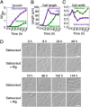
Magnesium sulfate treatment accelerated hyphal growth in T. asahii. (A) The growth rate, (B) length, and (C) width of JCM 2466 cells cultivated in Sabouraud medium (green) and Sabouraud medium containing 4.15 mM MgSO4 (purple) were measured at 8 h and every 24 h for 144 h at 25°C. The average length at each time point is given in panel B. The average width at each time point is shown in panel C. (D) The cell morphological phenotype of JCM 2466 cells is shown at each time point. The scale bar is 10 μm. OD660, optical density at 660 nm.
FIG 4
FIG 4
T. asahii cells retained the ability to form hyphae even after cultivation in Sabouraud medium. To determine whether JCM 2466 cells retain the ability to respond to MgSO4 addition after continuous cell culture in Sabouraud medium, cells were cultivated for 18 h at 25°C in Sabouraud medium, followed by further incubation for 20 h in Sabouraud+Mg medium (A) Scheme of culture of JCM 2466 cells in Sabouraud medium during 38 h. (B) Cell lengths after incubation for 18 and 38 h were measured and shown as the cell length classification. MgSO4, magnesium sulfate.
FIG 5
FIG 5
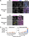
Large lipid droplets were resolved in hyphae of T. asahii. (A) Lipid droplets and DNA of JCM 2466 cells cultivated in Sabouraud medium and Sabouraud medium containing 4.15 mM MgSO4 for 16 h at 25°C were stained using Nile red and the DAPI, respectively. (B) Vacuoles in JCM 2466 cells were stained with FM4-64 after cultivation in Sabouraud medium and Sabouraud medium containing 4.15 mM MgSO4 for 16 h at 25°C. Yellow arrows indicate growing vacuoles. (C) The average area of all vacuoles in a cell was measured. Vacuoles with an area of ≥10 μm2 are indicated by a blue bar. Vacuoles with an area of <10 μm2 are indicated by an orange bar. Vacuolar area was classified based on cellular area. The numbers of cells in the respective size (area) ranges are as follows: in Sabouraud medium, 35 cells of 0 to 50 μm2, 17 cells of 50 to 100 μm2, 16 cells of 100 to 150 μm2, 12 cells of 150 to 200 μm2, 6 cells of 200 to 250 μm2, and 0 cells of greater than 250 μm2; in Sabouraud+Mg medium, 19 cells of 0 to 50 μm2, 13 cells of 50 to 100 μm2, 14 cells of 100 to 150 μm2, 14 cells of 150 to 200 μm2, 10 cells of 200 to 250 μm2, and 16 cells of greater than 250 μm2. The scale bar is 10 μm. DIC, differential interference contrast microscopy; YPD, yeast extract-peptone-dextrose medium.
FIG 6
FIG 6
Hyphal growth was inhibited in T. asahii by treatment with an actin inhibitor. (A) Lengths of JCM 2466 cells were measured after treatment with 100 μg/mL benzimidazole, 10 μg/mL latrunculin A, or DMSO (−) in YPD medium and Sabouraud medium containing 4.15 mM MgSO4 and then shown as the cell length classification. (B) Morphological phenotypes observed in experiments in panel A. The scale bar is 10 μm. BZ, benzimidazole; LA, latrunculin A; YPD, yeast extract-peptone-dextrose.
FIG 7
FIG 7
Mitochondria were distributed throughout the cell cytoplasm and adjacent to the cell walls in T. asahii and depended on magnesium supplementation. Mitochondria, lipid droplets, and the cell walls were stained with MitoBright, BODIPY, and calcofluor white, respectively, in JCM 2466 cells cultivated for 16 h at 25°C in YPD medium, Sabouraud medium, and Sabouraud medium containing 4.15 mM MgSO4. Hyphal cells are described in panel A, and yeast cells are described in panel B. Mito-C and Mito-S images indicate mitochondria distributions in the center and surface of a cell, respectively. The Mito-C images were used to produce the merged images. Arrows indicate the fragmented mitochondria in cells grown in Sabouraud medium. (C) Mitochondrial distribution was classified into following three phenotypes: distribution throughout the cell cytoplasm and adjacent to the cell walls (blue bar), fragmented distribution (orange bar), and reduced distribution (gray). The cell numbers in each size (length) range were as follows: in YPD medium: 32 cells of 0 to 10 μm, 134 cells of 10 to 20 μm, 92 cells of 20 to 30 μm, 20 cells of 30 to 40 μm, 5 cells of 40 to 50 μm, and 2 cells of greater than 50 μm; in Sabouraud medium, 182 cells of 0 to 10 μm, 53 cells of 10 to 20 μm, 19 cells of 20 to 30 μm, 4 cells of 30 to 40 μm, 2 cells of 40 to 50 μm, and 0 cells of greater than 50 μm; in Sabouraud+Mg medium, 80 cells of 0 to 10 μm, 49 cells of 10 to 20 μm, 34 cells of 20 to 30 μm, 24 cells of 30 to 40 μm, 21 cells of 40 to 50 μm, and 28 cells of greater than 50 μm.
FIG 8
FIG 8
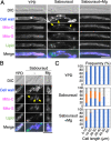
Mitochondrial distribution observed by confocal scanning microscopy. Mitochondria and vacuoles of JCM 2466 cells cultivated in (A) YPD medium, (B) Sabouraud medium, and (C) Sabouraud medium containing 4.15 mM MgSO4 for 16 h at 25°C were stained with MitoBright and FM4-64, respectively, and then observed under a confocal scanning microscope. White arrowheads indicate a vertical orange line showing the position of the cell cross section for the side view. A black arrowhead indicates a horizontal orange line showing the position of the hyphal cell cross-section for the side view. A hyphal form in Sabouraud medium is indicated by an arrow. A side view is shown of the hyphal form attached under the merged image. The dotted lines indicate a medial plane of a hyphal cell. The scale bar is 10 μm. YPD, yeast extract-peptone-dextrose; DIC, differential interference contrast microscopy.
FIG 9
FIG 9
Mitochondrial distribution was disrupted via treatment with actin inhibitor. JCM 2466 cells cultivated in (A) YPD medium, (B) Sabouraud medium, and (C) Sabouraud medium containing 4.15 mM MgSO4 for 16 h at 25°C were treated with 10 μg/mL latrunculin A or DMSO (−) and cultivated for 5 h at 25°C. Subsequently, mitochondria and lipid droplets were stained using MitoBright and BODIPY, respectively. NS, not-stained control. The scale bar is 10 μm. LA, latrunculin A; DIC, differential interference contrast microscopy; DMSO, dimethyl sulfoxide.
FIG 10
FIG 10
Model of the effects of magnesium on hyphal cell development. Shown are illustrations of T. asahii cells cultivated in Sabouraud medium with or without 4.15 mM MgSO4. In the cells cultivated in Sabouraud medium, there is an accumulation of large lipid droplets and fragmented mitochondria. In response to an increase in the concentration of Mg2+, T. asahii cells elongate and form hyphae. Within the hyphal cells, large lipid droplets are resolved, vacuoles are enlarged, and mitochondria are distributed throughout the cell cytoplasm and adjacent to the cell walls. V, vacuoles; LD, lipid droplets; Mito, mitochondria.

Similar articles

Cited by

References

    1. Boyce KJ, Andrianopoulos A. 2015. Fungal dimorphism: the switch from hyphae to yeast is a specialized morphogenetic adaptation allowing colonization of a host. FEMS Microbiol Rev 39:797–811. doi:10.1093/femsre/fuv035. - DOI - PubMed
    1. Jacobsen ID, Wilson D, Wächtler B, Brunke S, Naglik JR, Hube B. 2012. Candida albicans dimorphism as a therapeutic target. Expert Rev Anti Infect Ther 10:85–93. doi:10.1586/eri.11.152. - DOI - PubMed
    1. Berman J, Sudbery PE. 2002. Candida albicans: a molecular revolution built on lessons from budding yeast. Nat Rev Genet 3:918–930. doi:10.1038/nrg948. - DOI - PubMed
    1. Chen H, Zhou X, Ren B, Cheng L. 2020. The regulation of hyphae growth in Candida albicans. Virulence 11:337–348. doi:10.1080/21505594.2020.1748930. - DOI - PMC - PubMed
    1. Furuya K, Niki H. 2010. The DNA damage checkpoint regulates a transition between yeast and hyphal growth in Schizosaccharomyces japonicus. Mol Cell Biol 30:2909–2917. doi:10.1128/MCB.00049-10. - DOI - PMC - PubMed

Publication types

Supplementary concepts

LinkOut - more resources