. 2023 Apr 3;14(4):197.
doi: 10.3390/jfb14040197.

Lutein Encapsulated in PLGA-Phospholipid Nano-Carrier Effectively Mitigates Cytokines by Inhibiting Tumor Necrosis Factor TNF-α and Nuclear Factor NF- κ B in Mice Retina

Affiliations

Lutein Encapsulated in PLGA-Phospholipid Nano-Carrier Effectively Mitigates Cytokines by Inhibiting Tumor Necrosis Factor TNF-α and Nuclear Factor NF- κ B in Mice Retina

Ranganathan Arunkumar et al. J Funct Biomater. .

Abstract

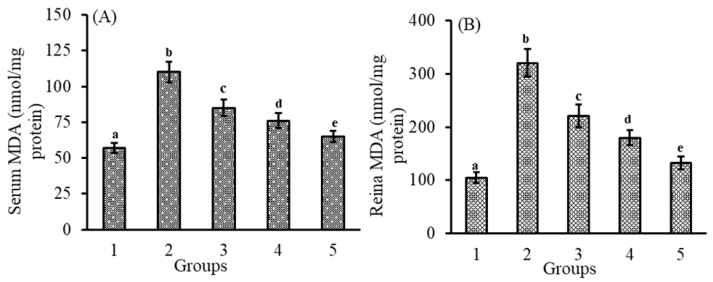
Lutein, a photo- and thermo-labile macular pigment, prevents the retina from suffering ocular inflammation with its antioxidant and anti-inflammatory activity. However, its biological activity is poor due to poor solubility and bioavailability. Therefore, we developed a PLGA NCs (+PL), (poly (lactic-co-glycolic acid) nanocarrier with phospholipid) to improve the biological availability and bioefficacy of lutein in the retina of lipopolysaccharide (LPS)-induced lutein-devoid (LD) mice. The effect of lutein-loaded NCs with/without PL was studied in comparison with micellar lutein. The induction of inflammation by LPS significantly increased the production of nitrites in the LPS-induced group, revealing higher levels of nitric oxide (NO) in the serum (760%) and retina (891%) compared to the control group. Malondialdehyde (MDA) levels in the serum (93%) and retina (205%) of the LPS-induced group were higher compared to the control group. LPS induction resulted in increased protein carbonyls in the serum (481%) and retina (487%) of the LPS group compared to the control group. Further, to conclude, lutein-PLGA NCs (+PL) effectively down-regulated inflammatory complications in the retina.

Keywords: LPS; PLGA; anti-inflammation; antioxidant; carotenoids; lutein; nanocarrier; phospholipids; retina.


Conflict of interest statement

There are no conflicts of interest to declare.

Figures

Figure 1
Figure 1
Effect of lutein-PLGA NCs (+PL)/(−PL) and micellar lutein on NO levels in serum (A) and retina (B) of LD mice previously induced with LPS. Groups: 1 = control, 2 = LPS, 3 = micellar lutein, 4 = lutein-PLGA NCs (−PL), and 5 = lutein-PLGA NCs (+PL) gavaged for 15 days. Values are mean ± SD (n = 6) and bars not sharing a similar superscript are significantly different (p < 0.05) among the groups.
Figure 2
Figure 2
Effect of lutein-PLGA NCs (+PL)/(−PL) and micellar lutein on MDA levels in serum (A) and retina (B) of LD mice previously induced with LPS. Groups: 1 = control, 2 = LPS, 3 = micellar lutein, 4 = lutein-PLGA NCs (−PL), and 5 = lutein-PLGA NCs (+PL) intubated for 15 days. Values are mean ± SD (n = 6) and bars not sharing a similar superscript are significantly different (p < 0.05) among the groups.
Figure 3
Figure 3
Effect of lutein-PLGA NCs (+PL)/(−PL) and micellar lutein on protein carbonyl levels in serum (A) and retina (B) of LD mice previously induced with LPS. Groups: 1 = control, 2 = LPS, 3 = micellar lutein, 4 = lutein-PLGA NCs (−PL), and 5 = lutein-PLGA NCs (+PL) intubated for 15 days. Values are mean ± SD (n = 6) and bars not sharing a similar superscript are significantly different (p < 0.05) among the groups.
Figure 4
Figure 4
Effect of lutein-PLGA NCs (+PL)/(−PL) and micellar lutein on TNF-α (A), MCP-1 (B), IL-6 (C), IL-1B (D), and PGE2 (E) levels in serum of LD mice induced with LPS. Groups: 1—Control, 2—LPS, 3—Micellar lutein, 4—Lutein-PLGA NCs (−PL) and 5—Lutein-PLGA NCs (+PL). Values are mean ± SD (n = 5) and bars not sharing similar superscript are significantly different (p < 0.05).
Figure 5
Figure 5
Protein expression pattern of iNOS (A) and COX-2 (C) in retina of LPS-induced LD mice. Lane 1—control, 2—LPS, 3—lutein-PLGA NCs (+PL), 4—lutein-PLGA NCs (-PL), 5—lutein. Relative values of iNOS (B) and COX-2 (D) over the control. Bars not sharing similar superscript are significantly different (p < 0.05).
Figure 6
Figure 6
Effect of lutein-PLGA NCs (+PL)/(−PL) and micellar lutein on NF-κB p65 subunit activation in retina of LPS-induced LD mice. 1 = control, 2 = LPS, 3 = micellar lutein, 4 = lutein-PLGA NCs (−PL), and 5 = lutein-PLGA NCs (+PL). Mean ± SD (n = 5) and bars not sharing similar superscript are significantly different (p < 0.05).
Figure 7
Figure 7
Bioavailability of lutein from lutein-PLGA NCs (+PL)/(−PL) and micellar lutein in the serum and retina of LPS-induced LD mice. Mean ± SD (n = 5) and bars not sharing similar superscript are significantly different (p < 0.05).

References

    1. Kauppinen A., Paterno J.J., Blasiak J., Salminen A., Kaarniranta K. Inflammation and Its Role in Age-Related Macular Degeneration. Cell Mol. Life Sci. 2016;73:1765–1786. doi: 10.1007/s00018-016-2147-8. - DOI - PMC - PubMed
    1. Rosenbaum J.T., McDevitt H.O., Guss R.B., Egbert P.R. Endotoxin-Induced Uveitis in Rats as a Model for Human Disease. Nature. 1980;286:611–613. doi: 10.1038/286611a0. - DOI - PubMed
    1. Fang H., Pengal R.A., Cao X., Ganesan L.P., Wewers M.D., Marsh C.B., Tridandapani S. Lipopolysaccharide-Induced Macrophage Inflammatory Response Is Regulated by SHIP. J. Immunol. 2004;173:360–366. doi: 10.4049/jimmunol.173.1.360. - DOI - PubMed
    1. Skrzypczak-Wiercioch A., Sałat K. Lipopolysaccharide-Induced Model of Neuroinflammation: Mechanisms of Action, Research Application and Future Directions for Its Use. Molecules. 2022;27:5481. doi: 10.3390/molecules27175481. - DOI - PMC - PubMed
    1. Hwang D., Kang M.-J., Jo M.J., Seo Y.B., Park N.G., Kim G.-D. Anti-Inflammatory Activity of β-Thymosin Peptide Derived from Pacific Oyster (Crassostrea gigas) on NO and PGE2 Production by Down-Regulating NF-ΚB in LPS-Induced RAW264.7 Macrophage Cells. Mar. Drugs. 2019;17:129. doi: 10.3390/md17020129. - DOI - PMC - PubMed