Review
. 2023 Mar 31;13(4):397.
doi: 10.3390/membranes13040397.

Extracellular Vesicles and Their Membranes: Exosomes vs. Virus-Related Particles

Affiliations
Review

Extracellular Vesicles and Their Membranes: Exosomes vs. Virus-Related Particles

Daniela Cortes-Galvez et al. Membranes (Basel). .

Abstract

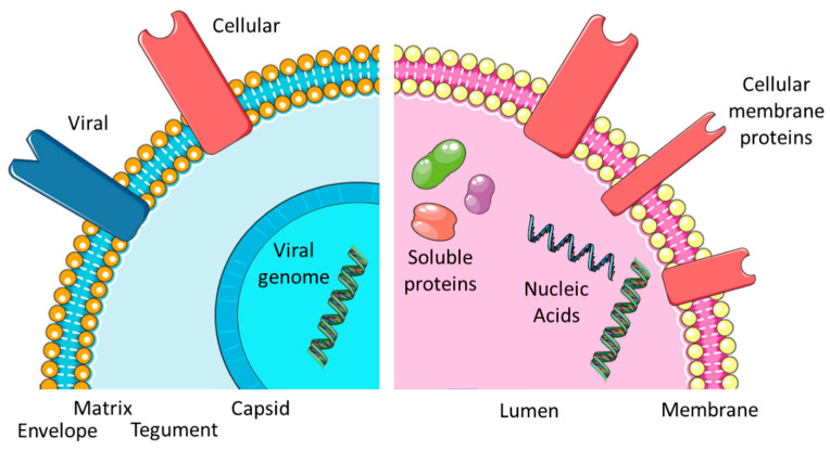
Cells produce nanosized lipid membrane-enclosed vesicles which play important roles in intercellular communication. Interestingly, a certain type of extracellular vesicle, termed exosomes, share physical, chemical, and biological properties with enveloped virus particles. To date, most similarities have been discovered with lentiviral particles, however, other virus species also frequently interact with exosomes. In this review, we will take a closer look at the similarities and differences between exosomes and enveloped viral particles, with a focus on events taking place at the vesicle or virus membrane. Since these structures present an area with an opportunity for interaction with target cells, this is relevant for basic biology as well as any potential research or medical applications.

Keywords: envelope; exosome; extracellular vesicle; membranes; vesicle; virus.


Conflict of interest statement

The authors declare no conflict of interest.

Figures

Figure 1
Figure 1
Comparing EXOs and EVIs. Overview of the virus and exosome morphology and content.
Figure 2
Figure 2
Comparing lifecycles. Both exosomes and virus particles transfer a package of signals from a producer cell to recipient cell. Cellular contribution (in green) and membrane functions (in orange) are similar, whereas initiating events (*) and perspectives (**) differ.
Figure 3
Figure 3
Biogenesis of exosomes and enveloped viruses. EXOs are produced from late endosomes (1) by the formation of interluminal vesicles (2), which are either released at the cell membrane to become exosomes (3) or recycled via fusion with lysosomes (4). Enveloped viruses acquire their envelope at different sites: nuclear envelope (A), endoplasmic reticulum or Golgi apparatus (B), followed by the release of exocytic vesicles containing virus particles (C). Alternatively, enveloped virus particles may acquire their envelope directly at the cell membrane (ectosomal budding) (D).

References

    1. Metzner C., Zaruba M. On the Interplay of Extracellular vesicles and Viral Infections. Trillium Extracell. Vesicles. 2020;1:12. doi: 10.47184/tev.2020.01.02. - DOI
    1. Zaruba M., Roschitz L., Sami H., Ogris M., Gerner W., Metzner C. Surface Modification of E. coli Outer Membrane Vesicles with Glycosylphosphatidylinositol-Anchored Proteins: Generating Pro/Eukaryote Chimera Constructs. Membranes. 2021;11:428. doi: 10.3390/membranes11060428. - DOI - PMC - PubMed
    1. Couch Y., Buzas E.I., Di Vizio D., Gho Y.S., Harrison P., Hill A.F., Lotvall J., Raposo G., Stahl P.D., Thery C., et al. A brief history of nearly EV-erything-The rise and rise of extracellular vesicles. J. Extracell. Vesicles. 2021;10:e12144. doi: 10.1002/jev2.12144. - DOI - PMC - PubMed
    1. Cocucci E., Meldolesi J. Ectosomes and exosomes: Shedding the confusion between extracellular vesicles. Trends Cell. Biol. 2015;25:364–372. doi: 10.1016/j.tcb.2015.01.004. - DOI - PubMed
    1. Record M., Silvente-Poirot S., Poirot M., Wakelam M.J.O. Extracellular vesicles: Lipids as key components of their biogenesis and functions. J. Lipid Res. 2018;59:1316–1324. doi: 10.1194/jlr.E086173. - DOI - PMC - PubMed

LinkOut - more resources