. 2023 Apr 10;9(2):810-828.
doi: 10.3390/tomography9020066.

Toward Practical Integration of Omic and Imaging Data in Co-Clinical Trials

Affiliations

Toward Practical Integration of Omic and Imaging Data in Co-Clinical Trials

Emel Alkim et al. Tomography. .

Abstract

Co-clinical trials are the concurrent or sequential evaluation of therapeutics in both patients clinically and patient-derived xenografts (PDX) pre-clinically, in a manner designed to match the pharmacokinetics and pharmacodynamics of the agent(s) used. The primary goal is to determine the degree to which PDX cohort responses recapitulate patient cohort responses at the phenotypic and molecular levels, such that pre-clinical and clinical trials can inform one another. A major issue is how to manage, integrate, and analyze the abundance of data generated across both spatial and temporal scales, as well as across species. To address this issue, we are developing MIRACCL (molecular and imaging response analysis of co-clinical trials), a web-based analytical tool. For prototyping, we simulated data for a co-clinical trial in "triple-negative" breast cancer (TNBC) by pairing pre- (T0) and on-treatment (T1) magnetic resonance imaging (MRI) from the I-SPY2 trial, as well as PDX-based T0 and T1 MRI. Baseline (T0) and on-treatment (T1) RNA expression data were also simulated for TNBC and PDX. Image features derived from both datasets were cross-referenced to omic data to evaluate MIRACCL functionality for correlating and displaying MRI-based changes in tumor size, vascularity, and cellularity with changes in mRNA expression as a function of treatment.

Keywords: breast cancer; cancer informatics; cancer modeling; magnetic resonance imaging (MRI); multi-omics; radiomics.


Conflict of interest statement

M.T.L. is founder of, and an uncompensated limited partner in, StemMed Ltd. and a founder of, and uncompensated manager in StemMed Holdings, its general partner. M.T.L. is also a founder of, and equity holder in, Tvardi Therapeutics Inc. L.E.D. is a compensated employee of StemMed Ltd. Some PDXs are exclusively licensed to StemMed Ltd., resulting in royalty income to L.E.D.

Figures

Figure 1
Figure 1
An illustrative overview of the flow of data and analysis within MIRACCL. The three columns describe the major components of data acquisition (blue), pre-processing (orange), and data analysis (green). The rows that cut across the three columns correspond to the clinical (top row) and pre-clinical settings (bottom row). Both DW-MRI and DCE-MRI are obtained for both settings, as are tissue samples. In the pre-processing stage, images are converted into quantitative parameter maps corresponding to the apparent diffusion coefficient (ADC, from DW-MRI) and the signal enhancement ratio (SER, from the DCE-MRI). Additionally, during this stage, the DNAseq (WES), RNAseq, and mass spectrometry proteomics are performed. The resulting data and images are then delivered to LinkedOmics and ePAD to summarize the output for subsequent input into MIRACCL.
Figure 2
Figure 2
Co-clinical trial design. (A) Overall RESPONSE clinical trial schema. For the purposes of this study, only the Stage T3 TNBC portion of the trial will be used in which patients will receive weekly paclitaxel (P) for a period of 12 weeks (four 21-day cycles) in combination with carboplatin (+/− pembrolizumab, p) given on day 1 of each of the four cycles. Imaging will be conducted at three timepoints. The first two timepoints will use MRI, while the third timepoint will use either ultrasound or mammography to assess response to the paclitaxel-containing regimens. Only the baseline biopsy is required in the trial; all others are optional. Our goal is to recruit 50 TNBC patients who will agree to have the first two biopsies for the generation of omic data (WES, RNAseq, MS-Proteomics). HER2+ patients will receive paclitaxel in combination with trastuzumab (T) and pertuzumab (P), while high risk ER+ patients will receive paclitaxel as a single agent. (B) PDX-based preclinical trial schema. Mice will receive four weekly cycles of combination paclitaxel/carboplatin with three MRI timepoints.
Figure 3
Figure 3
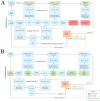
(A) Clinical workflow associated with a co-clinical trial. (B) Pre-clinical workflow associated with a co-clinical trial. The study workflows are diagramed centrally within each figure with specimen collection timepoints for omics generation shown above the study workflow and imaging collection timepoints shown below the workflow. Workflow steps shown in blue are standard across both the patient and PDX cohort while patient specific steps are shown in salmon and PDX specific steps in green.
Figure 4
Figure 4
Interactions between ePAD and MIRACCL. User activity within MIRACCL in steps one through four. Imaging data analysis API requests from MIRACCL to ePAD are shown on the top row while resulting MIRACCL user interface (UI) responses are shown on the bottom row.
Figure 5
Figure 5
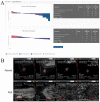
(A) Waterfall plot in MIRACCL of a treatment response in a cohort of patients (left) and in the corresponding cohort of PDX animals (right) based on the longest diameter of cancer lesions in pre-treatment and on-treatment images. These plots are generated by ePAD and displayed by MIRACCL. (B) Patient (top row) and corresponding PDX (bottom row) images as displayed in MIRACCL (tumor outlined in red). From left to right in the top row: T1W pre-treatment, T1W on-treatment, SER pre-treatment, and SER on-treatment for a patient from the I-SPY2 cohort. The bottom row shows four images of a biosimilar PDX model. From left to right: T1W pre-treatment, T1W on-treatment, SER pre-treatment, and SER on-treatment.
Figure 6
Figure 6
MIRACCL’s omics module. (A) The feature and dataset selector accepts user inputs which are passed to LinkedOmics via API for analysis. (A) Search results containing the top 500 and bottom 500 genes for patient and PDX are returned to MIRACCL and displayed in side-by-side tables. Venn diagrams visualizing the count of distinctive and overlapping genes between the patient and PDX cohorts are shown. (B) Volcano plots found on the Plots tab visually represent association results of Spearman tests for both patient and PDX cohorts.

References

    1. Petrosyan V., Dobrolecki L.E., Thistlethwaite L., Lewis A.N., Sallas C., Srinivasan R.R., Lei J.T., Kovacevic V., Obradovic P., Ellis M.J., et al. Identifying biomarkers of differential chemotherapy response in TNBC patient-Derived xenografts with a CTD/WGCNA approach. iScience. 2023;26:105799. doi: 10.1016/j.isci.2022.105799. - DOI - PMC - PubMed
    1. Shoghi K.I., Badea C.T., Blocker S.J., Chenevert T.L., Laforest R., Lewis M.T., Luker G.D., Manning H.C., Marcus D.S., Mowery Y.M., et al. Co-Clinical imaging resource program (CIRP): Bridging the translational divide to advance precision medicine. Tomography. 2020;6:273–287. doi: 10.18383/j.tom.2020.00023. - DOI - PMC - PubMed
    1. Ding L., Ellis M.J., Li S., Larson D.E., Chen K., Wallis J.W., Harris C.C., McLellan M.D., Fulton R.S., Fulton L.L., et al. Genome remodelling in a basal-Like breast cancer metastasis and xenograft. Nature. 2010;464:999–1005. doi: 10.1038/nature08989. - DOI - PMC - PubMed
    1. Li S., Shen D., Shao J., Crowder R., Liu W., Prat A., He X., Liu S., Hoog J., Lu C., et al. Endocrine-Therapy-Resistant ESR1 variants revealed by genomic characterization of breast-Cancer-Derived xenografts. Cell. Rep. 2013;4:1116–1130. doi: 10.1016/j.celrep.2013.08.022. - DOI - PMC - PubMed
    1. Koc S., Lloyd M.W., Grover J.W., Xiao N., Seepo S., Subramanian S.L., Ray M., Frech C., DiGiovanna J., Webster P., et al. PDXNet portal: Patient-Derived xenograft model, data, workflow and tool discovery. NAR Cancer. 2022;4:zcac014. doi: 10.1093/narcan/zcac014. - DOI - PMC - PubMed

Publication types

MeSH terms

LinkOut - more resources