. 2023 Apr 20;15(4):302.
doi: 10.3390/toxins15040302.

An Assassin's Secret: Multifunctional Cytotoxic Compounds in the Predation Venom of the Assassin Bug Psytalla horrida (Reduviidae, Hemiptera)

Affiliations

An Assassin's Secret: Multifunctional Cytotoxic Compounds in the Predation Venom of the Assassin Bug Psytalla horrida (Reduviidae, Hemiptera)

Maike Laura Fischer et al. Toxins (Basel). .

Abstract

Predatory assassin bugs produce venomous saliva that enables them to overwhelm, kill, and pre-digest large prey animals. Venom from the posterior main gland (PMG) of the African assassin bug Psytalla horrida has strong cytotoxic effects, but the responsible compounds are yet unknown. Using cation-exchange chromatography, we fractionated PMG extracts from P. horrida and screened the fractions for toxicity. Two venom fractions strongly affected insect cell viability, bacterial growth, erythrocyte integrity, and intracellular calcium levels in Drosophila melanogaster olfactory sensory neurons. LC-MS/MS analysis revealed that both fractions contained gelsolin, redulysins, S1 family peptidases, and proteins from the uncharacterized venom protein family 2. Synthetic peptides representing the putative lytic domain of redulysins had strong antimicrobial activity against Escherichia coli and/or Bacillus subtilis but only weak toxicity towards insect or mammalian cells, indicating a primary role in preventing the intake of microbial pathogens. In contrast, a recombinant venom protein family 2 protein significantly reduced insect cell viability but exhibited no antibacterial or hemolytic activity, suggesting that it plays a role in prey overwhelming and killing. The results of our study show that P. horrida secretes multiple cytotoxic compounds targeting different organisms to facilitate predation and antimicrobial defense.

Keywords: Reduviidae; cytotoxicity; redulysin; venom protein family 2; venomics.


Conflict of interest statement

The authors declare no conflict of interest.

Figures

Figure 1
Figure 1
SDS-PAGE analysis of fractions of Psytalla horrida PMG venom. 1–43 = venom fractions obtained through cation exchange chromatography; PMG = unfractionated PMG venom; M = protein marker. Fraction A and fraction B are highlighted in red. The first five fractions correspond to the flow-through with buffer A, whereas the last six fractions correspond to the flow-through with buffer B.
Figure 2
Figure 2
Bioactivity of fraction A and fraction B in comparison with a negative control (20 mM MES + 0.4 M NaCl or 20 mM MES + 1 M NaCl, respectively), and (+) a positive control treatment. Significant differences compared to the respective negative control are highlighted with asterisks (* p ≤ 0.05; ** p ≤ 0.01; *** p ≤ 0.001; ns = not significant). (A) Fraction A led to reduced viability of treated sf9 cells; (+) = 0.1% Triton x-100. Statistical test: Dunn’s tests, n = 6. (B) Both fractions significantly delayed the growth of Escherichia coli; (+) = 0.05 mg/mL gentamycin. Statistical test: pairwise t-tests, n = 3. (C) Fraction B caused mild hemolysis of horse erythrocytes; (+) = 0.1% Triton x-100. Statistical test: Dunn’s tests, n = 3.
Figure 3
Figure 3
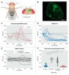
Calcium imaging of Drosophila melanogaster antennal lobes using genetic expression of the calcium-sensitive protein GCaMP6s. (A) Schematic of Drosophila melanogaster highlighting the left antennal lobe that was imaged. (B) Representative image of an antennal lobe with GCaMP6s expression in olfactory sensory neurons without venom treatment. The dashed line represents the region selected with the ROI manager to extract the brightness values. (C) Changes of dF/F (representing fluorescence changes as an indicator for intracellular calcium concentration) after treatment with unfractionated PMG venom; for highlighting the observed representative course of the fluorescence change, one replicate is marked in dark red. (D) Changes of dF/F after treatment with fraction A. (E) Changes of dF/F after treatment with 20 mM MES and 20 mM MES + 0.6 M NaCl (negative controls). (F) Changes in fluorescence intensity after 10 min. Boxplots within the violin plots represent the median (line), interquartile range (box), and data range (whiskers). Significant differences between treatments are highlighted with asterisks (** p ≤ 0.01; *** p ≤ 0.001, Dunn’s tests, n = 10 (PMG, fraction A, 20 mM MES + 0.6 M NaCl), n = 11 (20 mM MES)).
Figure 4
Figure 4
Protein composition of fraction A and fraction B. Color-coded blocks represent the number of contigs identified in transcriptome datasets and verified by proteomic analysis, which encodes specific classes of functional proteins. Log2(Total Score) depicts the logarithmic total score of all matched peptides identified by LC-MS/MS. Log2(RPKM) shows the expression level of the respective contig in the PMG.
Figure 5
Figure 5
Bioactivity of synthetic redulysin peptides. (A) The structure of redulysins consists of a signal peptide, a proregion, a lytic region, and a non-lytic region. After cleavage of the proregion at the DEER motif, the amphipathic pore-forming alpha-helix is exposed. (B) Insect cell viability, erythrocyte integrity and growth of Escherichia coli, Bacillus subtilis and Bacillus thuringiensis in presence of 10 µM redulysin peptide; (−) = 20 mM MES pH 5.5 (=100%), (+) = 0.5 mg/mL gentamycin or 0.1% triton-x 100. Significant differences compared to the negative control are highlighted with asterisks (* p ≤ 0.05; ** p ≤ 0.01; *** p ≤ 0.001, pairwise t-tests or Dunn’s tests, n = 3). (C) Dose-dependent growth of Escherichia coli 14 h after treatment with selected redulysin peptides at varying concentrations. The data were fitted to a logistic model and plotted as dose-response curves (n = 2). (D) Change of intracellular Ca2+ levels (represented by dF/F) in Drosophila melanogaster olfactory sensory neurons after treatment with 100 µM redulysin peptide 18. Violin plots represent the change in fluorescence intensity after 10 min (* p ≤ 0.05, Kruskal–Wallis test). Boxplots within the violin plots represent the median (line), interquartile range (box), and data range (whiskers). (−) = 20 mM MES pH 5.5. Peptide 18, which corresponds to the redulysin from the phytozoophagous bug Lygus rugulipennis is marked with a leaf symbol in (BD).
Figure 6
Figure 6
Bioactivity of a recombinant venom protein family 2 (Vpf2) protein. (A) Insect cell viability in the presence of 0.7 mg/mL Vpf2 and heated Vpf2; (−) = PBS (=100%), (+) = 0.1% triton-x 100. Significant differences compared to the negative control are highlighted with asterisks (* p ≤ 0.05; *** p ≤ 0.001; ns = not significant, Dunn’s tests, n = 5). (B) Course of intracellular Ca2+ levels in Drosophila melanogaster olfactory neurons after treatment with 0.2 mg/mL Vpf2 or PBS. Violin plots represent the change in fluorescence intensity after 10 min (ns = not significant; α = 0.05, Kruskal-Wallis test). Boxplots within the violin plots represent the median (line), interquartile range (box), and data range (whiskers).

References

    1. Walker A.A., Madio B., Jin J., Undheim E.A., Fry B.G., King G.F. Melt with This Kiss: Paralyzing and Liquefying Venom of the Assassin Bug Pristhesancus plagipennis (Hemiptera: Reduviidae) Mol. Cell. Proteom. 2017;16:552–566. doi: 10.1074/mcp.M116.063321. - DOI - PMC - PubMed
    1. Ables J. Feeding behavior of an assassin bug, Zelus renardii. Ann. Entomol. Soc. Am. 1978;71:476–478. doi: 10.1093/aesa/71.4.476. - DOI
    1. Dos Santos C.E.P., de Souza J.R., Zanette R.A., da Silva F.J., Strussmann C. Bite caused by the assassin bug Zelus Fabricius, 1803 (Hemiptera; Heteroptera: Reduviidae) in a human. Wilderness Environ. Med. 2019;30:63–65. doi: 10.1016/j.wem.2018.10.002. - DOI - PubMed
    1. Hartwig E. Records of assassin bug species (reduviidae, heteroptera) reported biting man. Koedoe. 1977;20:109–114. doi: 10.4102/koedoe.v20i1.935. - DOI
    1. Walker A.A., Mayhew M.L., Jin J., Herzig V., Undheim E.A.B., Sombke A., Fry B.G., Meritt D.J., King G.F. The assassin bug Pristhesancus plagipennis produces two distinct venoms in separate gland lumens. Nat. Commun. 2018;9:755. doi: 10.1038/s41467-018-03091-5. - DOI - PMC - PubMed

Publication types

LinkOut - more resources