. 2023 Apr 28;380(6643):387-391.
doi: 10.1126/science.adg9829. Epub 2023 Apr 27.

Bacterial spore germination receptors are nutrient-gated ion channels

Affiliations

Bacterial spore germination receptors are nutrient-gated ion channels

Yongqiang Gao et al. Science. .

Abstract

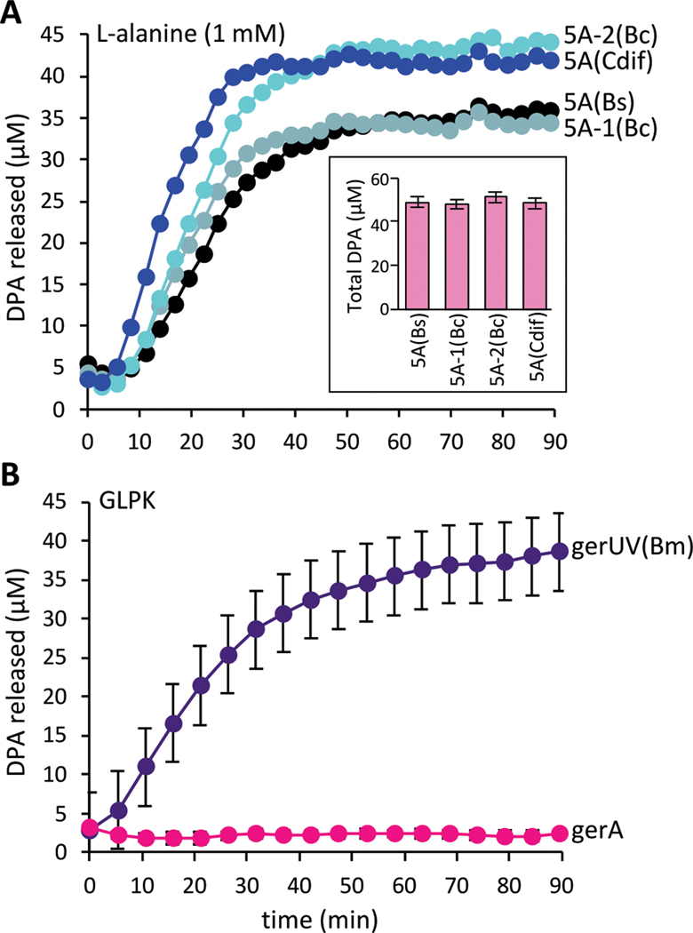
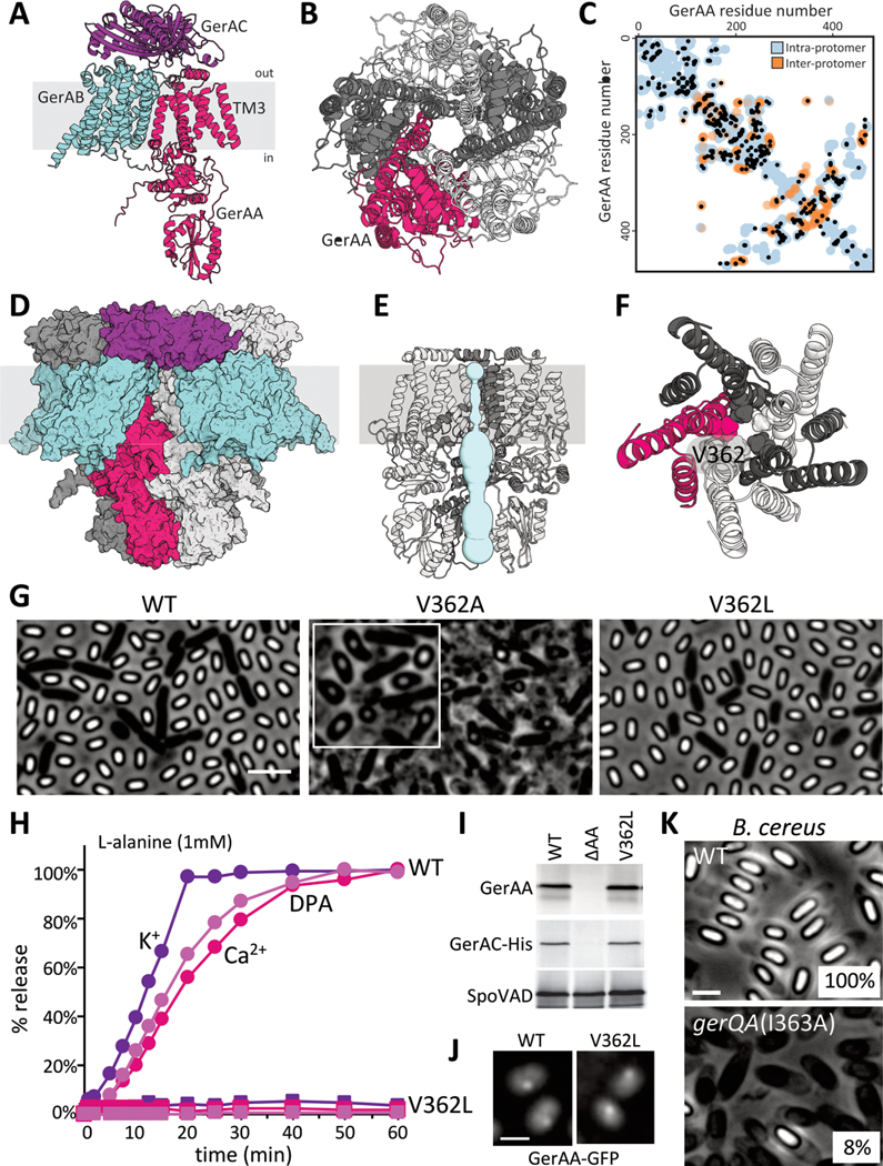
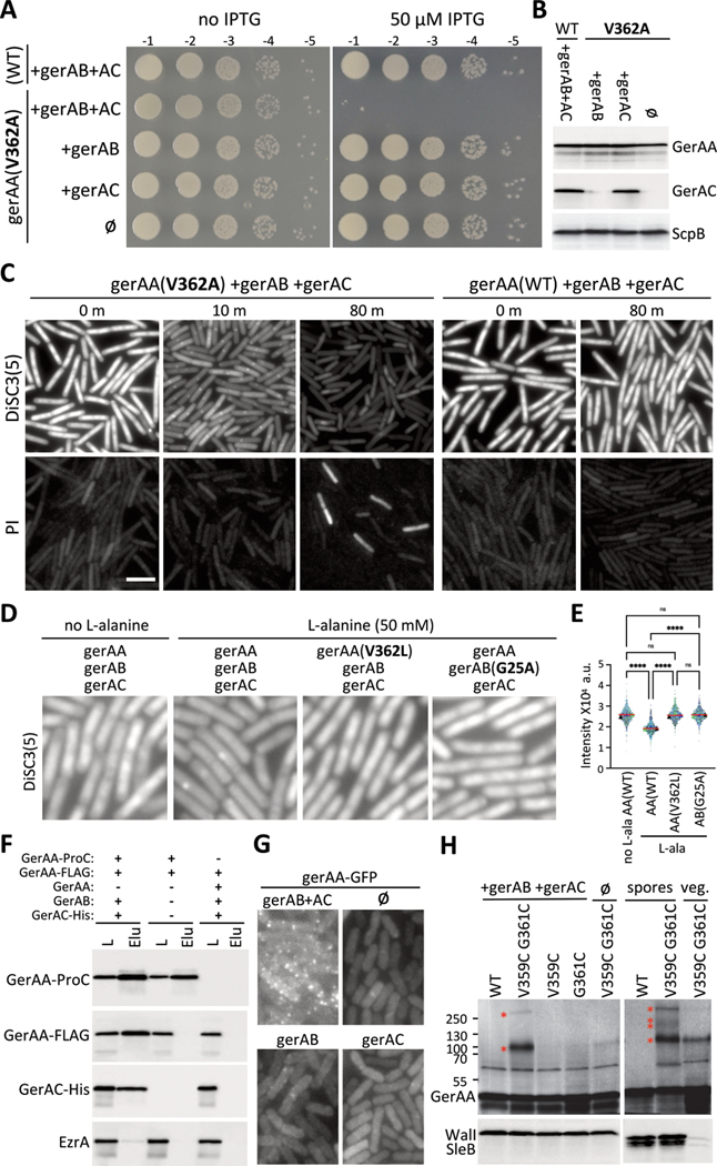
Bacterial spores resist antibiotics and sterilization and can remain metabolically inactive for decades, but they can rapidly germinate and resume growth in response to nutrients. Broadly conserved receptors embedded in the spore membrane detect nutrients, but how spores transduce these signals remains unclear. Here, we found that these receptors form oligomeric membrane channels. Mutations predicted to widen the channel initiated germination in the absence of nutrients, whereas those that narrow it prevented ion release and germination in response to nutrients. Expressing receptors with widened channels during vegetative growth caused loss of membrane potential and cell death, whereas the addition of germinants to cells expressing wild-type receptors triggered membrane depolarization. Therefore, germinant receptors act as nutrient-gated ion channels such that ion release initiates exit from dormancy.


Conflict of interest statement

Competing interests:

The authors declare no competing interests. DSM is a cofounder of Seismic Therapeutics, an advisor for Dyno Therapeutics, Octant, Jura Bio, Tectonic Therapeutics and Genentech.

Figures

Figure 1
Figure 1. Cross-species complementation of key germination factors.
(A) spoVA loci from B. cereus and C. difficile support DPA release from B. subtilis spores in response to L-alanine. Purified spores of ΔspoVA mutant strains harboring an ectopic copy of the indicated spoVA (5A) locus from B. subtilis (Bs), B. cereus (Bc), or C. difficile (Cdif). Spores were mixed with 1 mM L-alanine and DPA release was monitored over time. The insert shows total DPA content in purified spores. Representative data from one of three biological replicates are shown. The other two replicates can be found in Figure S2. (B) B. subtilis spores harboring the gerUV locus from B. megaterium germinate in response to D-glucose, L-leucine, L-proline and K+ (GLPK). Purified B. subtilis spores lacking all 5 endogenous germinant receptor loci (Δ5) and harboring the gerUV or gerA locus were incubated with GLPK (10 mM each) and DPA release was monitored over time. The data represent the average results from three biological replicates. Error bars are the standard deviations from the means. Similar results were obtained using a germination assay that monitors the drop in optical density as phase-bright spores transition to phase-dark (Fig. S3 and S4).
Figure 2.
Figure 2.. Evidence that GerAA forms a membrane channel.
(A) Predicted structure of the GerAA (red), GerAB (cyan), GerAC (purple) trimer. Topology is based on protease accessibility studies of GerAC and GerAA (10, 38). TM3, the lumen-adjacent helix in GerAA is labeled. (B) Predicted GerAA pentamer as viewed from outside the spore. Protomers are shown in dark and light gray and red. (C) Evolutionarily coupled (EC) residue pairs in GerAA are plotted as black circles. Intra-protomer (blue circles) and inter-protomer (orange circles) residue pairs that are ≤5 Å apart in the predicted GerAA pentamer are shown. (D) Space-filling model of the predicted pentamer of trimers. (E) Predicted pore (light blue) in the GerAA pentamer. Only three GerAA protomers are shown for clarity. (F) Top view of the GerAA hexamer model showing the concentric TM rings surrounding the channel. V362 is highlighted. (G) Representative phase-contrast images of sporulated cultures of strains harboring a second copy of gerAA(WT) or gerAA(V362A). The strain harboring gerAA(V362L) lacks the native gerAA copy. Scale bar, 3 μm. Inset highlights the teardrop-shaped spores in the V362A mutant. (H) Purified spores that have GerAA(WT) (circles) or GerAA(V362L) (squares) as the sole copy of the GerAA subunit were mixed with 1 mM L-alanine and the germination exudates were analyzed for K+, Ca2+, and DPA over time. (I) Immunoblots from lysates of the purified spores used in (H). GerAA(WT) and GerAA(V362L) are stable and stabilize GerAC-His, unlike spores lacking GerAA (ΔAA). SpoVAD controls for loading. (J) Representative fluorescence images of GerAA(WT)- and GerAA(V362L)-GFP localization in spores. Both localize in germinosome foci. Scale bar, 3 μm. (K) Representative phase-contrast images of sporulated cultures of wild-type B. cereus and a merodiploid strain harboring gerQA(I363A). Sporulation efficiency of each strain is indicated in the bottom right. Scale bar, 2μm. Representative data from one of at least three biological replicates are shown for (G), (H), (J), (K), and from one of two biological replicates for (I).
Figure 3.
Figure 3.. The GerA complex behaves like a nutrient-gated ion channel when expressed in vegetatively growing cells.
(A) Serial dilutions of the indicated strains with IPTG-regulated gerAA(WT) and gerAA(V362A) alleles and constitutively expressed gerAB and gerAC (AC). (B) Immunoblot analysis of the strains in (A). GerAA(WT) and GerAA(V362A) were expressed at similar levels in the presence or absence of GerAB and GerAC. ScpB controls for loading. (C) Representative fluorescence images of exponentially growing cultures of the indicated strains from (A). Time (in min) after IPTG addition is indicated. The top panels show fluorescence of the potentiometric dye DiSC3(5). The lower panels show propidium iodide (PI) staining. The two fields are from the same culture but stained and imaged separately. Scale bar, 5 μm. (D) Representative DiSC3(5) fluorescence images of exponentially growing cultures of the indicated strains 30 min after addition of 50 mM L-alanine. gerAA and gerAA(V362L) are IPTG-regulated alleles and gerAB, gerAB(G25A) and gerAC were expressed constitutively. (E) Quantitative analysis of DiSC3(5) fluorescence intensity from the same strains and conditions as in (D). DiSC3(5) fluorescence intensities were quantified from three biological replicates (>500 cells for each) and plotted in different colors. Triangles represent the median fluorescence intensity for each replicate, red lines show the median values for all cells per strain. P-value <0.0001 (****) and not significant (ns) are indicated. (F) Immunoblots of anti-ProC immuno-affinity purifications from detergent-solubilized membrane preparations of vegetatively growing B. subtilis cells expressing the indicated proteins. Load (L) and elution (Elu) are shown. GerAA-FLAG co-purifies with GerAA-ProC, provided GerAB and GerAC are co-expressed. The membrane protein EzrA serves as a negative control. (G) Representative fluorescence images of vegetative cells expressing GerAA-GFP in the presence and absence of GerAB and GerAC (AC). (H) Immunoblots of vegetative cells expressing cysteine-substituted GerAA variants in the presence or absence of GerAB and GerAC. GerAA(V359C G361C) produces disulfide species (red asterisks) with sizes of dimer and pentamer (left). WalI controls for loading. GerAA species of similar size were also detected from spore lysates (right). Two additional species were also detected. SleB controls for loading. Representative data from one of at least three biological replicates are shown for (A), (C-E), (G) and (H). (B) and (F) are from one of two biological replicates.

Comment in

References

    1. Andre S, Vallaeys T, Planchon S, Spore-forming bacteria responsible for food spoilage. Res Microbiol 168, 379–387 (2017). - PubMed
    1. Mallozzi M, Viswanathan VK, Vedantam G, Spore-forming Bacilli and Clostridia in human disease. Future Microbiol 5, 1109–1123 (2010). - PubMed
    1. Setlow P, Spore Resistance Properties. Microbiol Spectr 2, (2014). - PubMed
    1. Moir A, Cooper G, Spore Germination. Microbiol Spectr 3, (2015). - PubMed
    1. Setlow P, Wang S, Li YQ, Germination of Spores of the Orders Bacillales and Clostridiales. Annu Rev Microbiol 71, 459–477 (2017). - PubMed

MeSH terms

LinkOut - more resources