. 2023:38:103409.
doi: 10.1016/j.nicl.2023.103409. Epub 2023 Apr 19.

Distinct and joint effects of low and high levels of Aβ and tau deposition on cortical thickness

Affiliations

Distinct and joint effects of low and high levels of Aβ and tau deposition on cortical thickness

Seyed Hani Hojjati et al. Neuroimage Clin. 2023.

Abstract

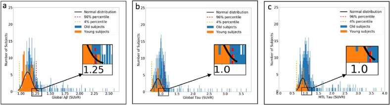
Alzheimer's disease (AD) is defined by the presence of Amyloid-β (Aβ),tau, and neurodegeneration (ATN framework) in the human cerebral cortex. Yet, prior studies have suggested that Aβ deposition can be associated with both cortical thinning and thickening. These contradictory results are attributed to small sample sizes, the presence versus absence of tau, and limited detectability in the earliest phase of protein deposition, which may begin in young adulthood and cannot be captured in studies enrolling only older subjects. In this study, we aimed to find the distinct and joint effects of Aβ andtau on neurodegeneration during the progression from normal to abnormal stages of pathologies that remain elusive. We used18F-MK6240 and 18F-Florbetaben/18F-Florbetapir positron emission tomography (PET) and magnetic resonance imaging (MRI) to quantify tau, Aβ, and cortical thickness in 590 participants ranging in age from 20 to 90. We performed multiple regression analyses to assess the distinct and joint effects of Aβ and tau on cortical thickness using 590 healthy control (HC) and mild cognitive impairment (MCI) participants (141 young, 394 HC elderlies, 52 MCI). We showed thatin participants with normal levels of global Aβdeposition, Aβ uptakewassignificantly associated with increasedcortical thickness regardless of tau (e.g., left entorhinal cortex with t > 3.241, p < 0.0013). The relationship between tau deposition and neurodegeneration was more complex: in participants with abnormal levels of global tau, tau uptake was associated with cortical thinning in several regions of the brain (e.g., left entorhinal with t < -2.80, p < 0.0096 and left insula with t-value < -4.284, p < 0.0001), as reported on prior neuroimaging and neuropathological studies. Surprisingly, in participants with normal levels of global tau, tau was found to be associated with cortical thickening. Moreover, in participants with abnormal levels of global Aβandtau, theresonancebetween them, defined as their correlation throughout the cortex, wasassociated strongly with cortical thinning even when controlling for a direct linear effect. We confirm prior findings of an association between Aβ deposition and cortical thickening and suggest this may also be the case in the earliest stages of deposition in normal aging. We also illustrate that resonance between high levels of Aβ and tau uptake is strongly associated with cortical thinning, emphasizing the effects of Aβ/tau synergy inAD pathogenesis.

Keywords: Alzheimer's disease; Amyloid-β; Cortical thickness; Neurodegeneration; Tau.


Conflict of interest statement

Declaration of Competing Interest The authors declare that they have no known competing financial interests or personal relationships that could have appeared to influence the work reported in this paper.

Figures

Fig. 1
Fig. 1
Overall procedures of this study.
Fig. 2
Fig. 2
Distribution of younger (in orange) and older (in blue) participants’, (a) global Aβ, (b) global tau, and (c) MTL tau. The fitted normal distribution (in black), 96th percentile (red dotted line), and 4th percentile (yellow dotted line) overlaid on the distributions of the participants. (For interpretation of the references to color in this figure legend, the reader is referred to the web version of this article.)
Fig. 3
Fig. 3
(a) Illustrating the vertex-wise probabilistic atlas of Aβ (left column) and tau (right column) pathologies throughout the entire cerebral cortex obtained in four participant categories. (First row, nAβ/nTau; second row, aAβ/nTau; third row, nAβ/aTau; and fourth row, aAβ/aTau). The probability of observing Aβ and tau at each vertex is color-coded with a heat color map and overlaid on a semi-inflated cortical surface of the MNI152 template. (b) Boxplots compare the distribution of global Aβ in four categories of participants. (c) Boxplots compare the distribution of global tau in four participant categories.
Fig. 4
Fig. 4
Inter-regional cross-correlogram between Aβ and Tau accumulations for (a) nAβ/nTau, (b) aAβ/nTau; (c) nAβ/aTau, and (d) aAβ/aTau groups in 76 cortical and subcortical regions. The correlation is color-coded with a heatmap, and the red color indicates a correlation value equal to 1, and the blue color indicates a correlation equal to −1. (For interpretation of the references to color in this figure legend, the reader is referred to the web version of this article.)
Fig. 5
Fig. 5
Vertex-wise statistical map (t-value) of association between global Aβ (left column), global tau (right column) pathologies, and cortical thickness throughout the entire cerebral cortex obtained in four categories of participants. (First row, nAβ/nTau; second row, aAβ/nTau; third row, nAβ/aTau; and fourth row, aAβ/aTau). The t-value at each vertex is color-coded with red to yellow colors representing increasing positive t-values and blue to light blue representing decreasing negative t-values and overlaid on the semi-inflated cortical surface of the MNI152 template. The association between global Aβ, global tau pathologies, and cortical thickness survived after multiple comparison corrections with FDR. (For interpretation of the references to color in this figure legend, the reader is referred to the web version of this article.)
Fig. 6
Fig. 6
(a) Region-wise statistical map (t-value) of association between regional Aβ (left column), regional tau (right column) pathologies, and regional cortical thickness throughout 68 cortical ROIs obtained in four categories of participants (First row, nAβ/nTau; second row, aAβ/nTau; third row, nAβ/aTau; and fourth row, aAβ/aTau). The t-value at each region is color-coded with red to yellow colors representing increasing positive t-values and blue to light blue representing decreasing negative t-values and overlaid on the semi-inflated cortical surface of the MNI152 template. (b) The regional multiple regression analysis results in the association between residual regional cortical thickness and Aβ in two target regions: left entorhinal and right posterior cingulate. (c) The regional multiple regression analysis results of the association between residual regional cortical thickness and tau, in two target regions: left entorhinal, and left insula. Association between regional Aβ, tau pathologies and cortical thickness survived after family-wise error correction. In Fig. 6b-c the survived associations relationships are shown with thicker lines and *. (For interpretation of the references to color in this figure legend, the reader is referred to the web version of this article.)
Fig. 7
Fig. 7
(a) Vertex-wise statistical map (t-value) of the association between resonance and cortical thickness throughout the entire cerebral cortex obtained in four categories of participants in aAβ/aTau group. The t-value at each vertex is color-coded with a heatmap where red to yellow shading represents increasing positive t-values and blue to light-blue shading represents decreasing negative t-values overlaid on the semi-inflated cortical surface of the MNI152 template. Association between resonance and cortical thickness survived after multiple comparison corrections with FDR. (For interpretation of the references to color in this figure legend, the reader is referred to the web version of this article.)
Fig. 8
Fig. 8
(a) Region-wise statistical map (t-value) of the association between resonance and cortical thickness throughout 68 cortical ROIs obtained in aAβ/aTau group. The t-value at each region is color-coded with a heatmap where red to yellow colors represent increasing positive t-values and blue to light-blue represents decreasing negative t-values overlaid on the semi-inflated cortical surface of the MNI152 template. (b) The regional multiple regression analysis results demonstrate the association between regional cortical thickness and resonance in two target regions: left parahippocampal, and right caudal anterior cingulate. Association between resonance and cortical thickness survived after family-wise error correction. In Fig. 8b the survived associations relationships are shown with thicker lines and *. (For interpretation of the references to color in this figure legend, the reader is referred to the web version of this article.)

References

    1. Ashrafian H., et al. Review on Alzheimer's disease: inhibition of amyloid beta and tau tangle formation. Int. J. Biol. Macromol. 2021;167:382–394. - PubMed
    1. Avants B.B., et al. Advanced normalization tools (ANTS) Insight j. 2009;2:1–35.
    1. Batzu L., et al. Cerebrospinal fluid progranulin is associated with increased cortical thickness in early stages of Alzheimer's disease. Neurobiol. Aging. 2020;88:61–70. - PubMed
    1. Becker J., et al. Amyloid deposition and brain volume across the continuum of aging and AD. Ann Neurol. 2011
    1. Braak H., et al. Staging of Alzheimer disease-associated neurofibrillary pathology using paraffin sections and immunocytochemistry. Acta Neuropathol. 2006;112:389–404. - PMC - PubMed