. 2023 Apr 27;14(1):2421.
doi: 10.1038/s41467-023-38160-x.

DNA polymerase POLD1 promotes proliferation and metastasis of bladder cancer by stabilizing MYC

Affiliations

DNA polymerase POLD1 promotes proliferation and metastasis of bladder cancer by stabilizing MYC

Yejinpeng Wang et al. Nat Commun. .

Abstract

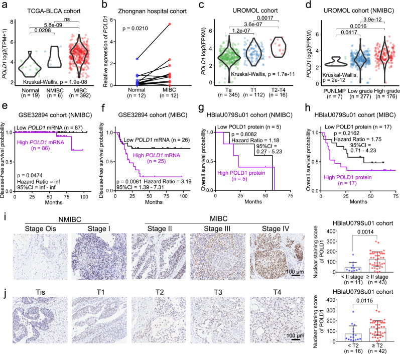
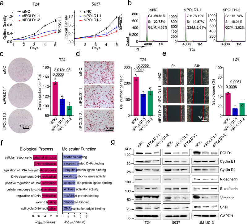
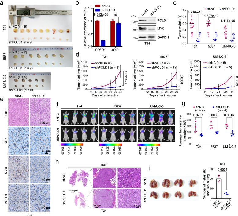
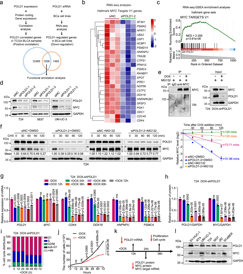
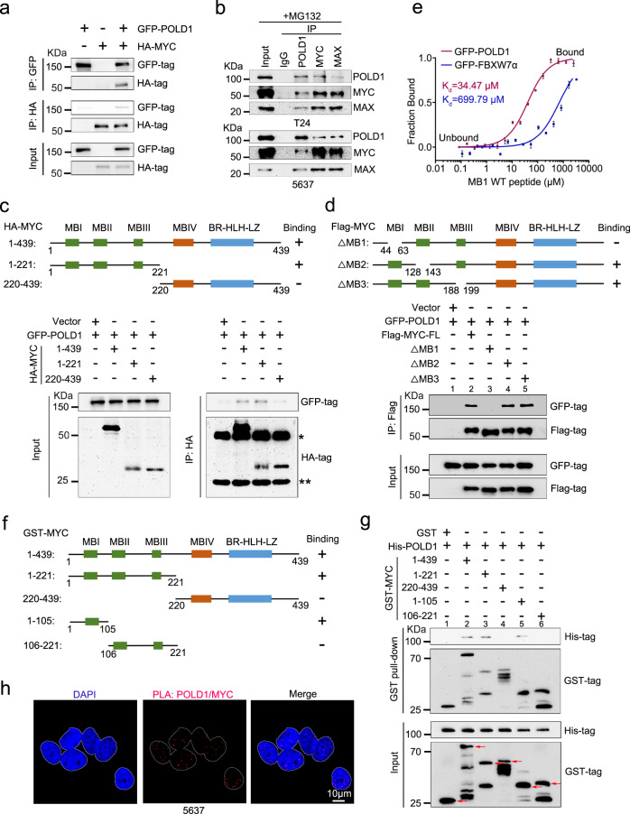
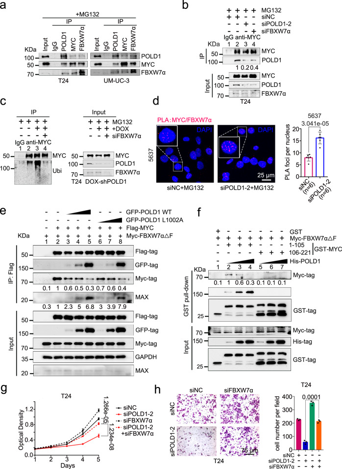
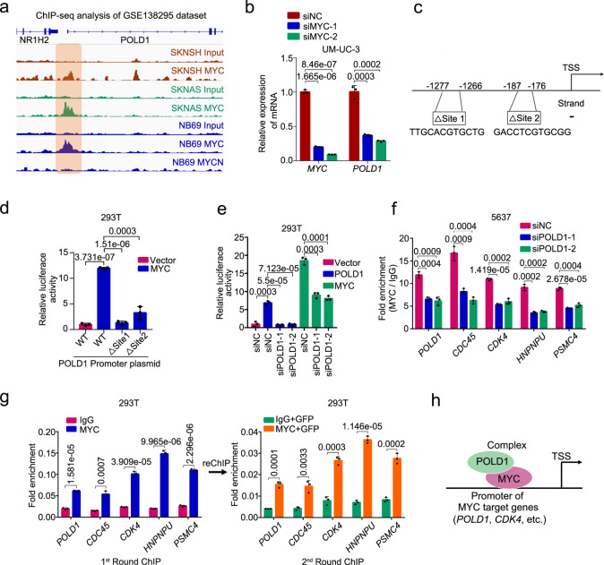
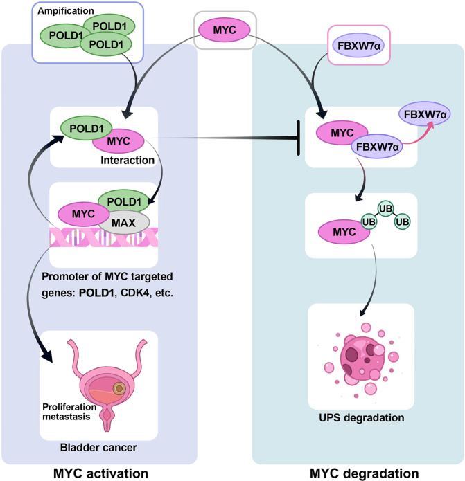
To date, most studies on the DNA polymerase, POLD1, have focused on the effect of POLD1 inactivation mutations in tumors. However, the implications of high POLD1 expression in tumorigenesis remains elusive. Here, we determine that POLD1 has a pro-carcinogenic role in bladder cancer (BLCA) and is associated to the malignancy and prognosis of BLCA. Our studies demonstrate that POLD1 promotes the proliferation and metastasis of BLCA via MYC. Mechanistically, POLD1 stabilizes MYC in a manner independent of its' DNA polymerase activity. Instead, POLD1 attenuates FBXW7-mediated ubiquitination degradation of MYC by directly binding to the MYC homology box 1 domain competitively with FBXW7. Moreover, we find that POLD1 forms a complex with MYC to promote the transcriptional activity of MYC. In turn, MYC increases expression of POLD1, forming a POLD1-MYC positive feedback loop to enhance the pro-carcinogenic effect of POLD1-MYC on BLCA. Overall, our study identifies POLD1 as a promotor of BCLA via a MYC driven mechanism and suggest its potential as biomarker for BLCA.


Conflict of interest statement

The authors declare no competing interests.

Figures

Fig. 1
Fig. 1. The expression of POLD1 was correlated with the malignant degree and prognosis of BLCA.
The mRNA level of POLD1 in BLCA (MIBC or NMIBC) and normal tissues in a TCGA-BLCA (RNA-seq data) and b Zhongnan Hospital cohort (RT-qPCR data). Statistical significance was determined by two-tailed Wilcoxon rank-sum test (a) and paired two-tailed Student’s T-test (b). The mRNA level of POLD1 in different c stages (Ta, T1, T2, T3, and T4) and d pathology grades: Papillary Urothelial Neoplasms of Low Malignant Potential (PUNLMP), low grade and high grade in the UROMOL cohort. Statistical significance was determined by two-tailed Wilcoxon rank-sum test and Kruskal–Wallis test (c, d). The prognostic curve (disease-free survival) of different POLD1 mRNA levels in the two subtypes of e NMIBC and f MIBC in the GSE32894 cohort was analyzed. The patients were divided into a high POLD1 mRNA level group and a low POLD1 mRNA level group according to the median POLD1 expression. The prognostic curve (overall survival) of different POLD1 protein levels in the two subtypes of g NMIBC and h MIBC in the HBlaU079Su01 cohort was analyzed. The patients were divided into high and low POLD1 protein level groups according to the median POLD1 protein level. Statistical significance was determined by the log-rank test of Kaplan–Meier analysis (eh). The purple line represents the group with high POLD1 mRNA or protein expression and the black line represents the group with low POLD1 mRNA or protein expression. Representative pictures (left panel) and statistical figures (right panel) of immunohistochemistry staining analysis of POLD1 protein levels in different i stages (The seventh edition of AJCC: stage Ois, stage I, stage II, stage III, and stage IV), and j T stages (Tis, T1, T2, T3, and T4) in the HBlaU079Su01 cohort. POLD1 nuclear staining score = The intensity of nuclear staining × Staining positive area. Data are mean ± SD. Statistical significance was determined by the two-tailed Student’s T-test (i, j). The n number represents n biologically independent patient samples in each group. Exact n values are marked in the images. NMIBC non-muscle-invasive bladder cancer, MIBC muscle-invasive bladder cancer, CI confidence interval, inf infinite, TPM transcripts per kilobase million, FPKM fragments per kilobase million. Source data are provided as a Source Data file; ns: not significant.
Fig. 2
Fig. 2. POLD1 promotes BLCA proliferation and metastasis in vitro.
a The cell proliferation curve of T24 and 5637 cells with POLD1 knockdown. Before detecting the absorbance, 20 μL of MTT was added to the 96-well seeded with cells, and incubated in a 37°C cell incubator for 4 h. Then, 200 μL of DMSO was added to each well and shaken until homogenized. Graph shows mean ± SD, n = 6 biologically independent experiments in each group. b Flow cytometric analysis of DNA content for cell cycle progression from propidium iodide (PI, 100 μg/mL) staining of T24 cells with or without POLD1 depletion. c Representative images (left panel) and statistical graph (right panel) of clone formation assays from the indicated groups with POLD1 depletion in T24 cells (n = 4 biologically independent experiments in each group). d Representative images (left panel) and cell numbers (right panel) of Transwell assays from the indicated groups with or without POLD1 knockdown in T24 cells (n = 3 biologically independent experiments in each group). e Representative images (left panel) and statistical results (right panel) of the wound healing assay in T24 cells with or without POLD1 depletion (n = 3 biologically independent experiments in each group). Gap closure (%) = (0 h distance − 24 h distance)/0 h distance 100%. The “distance” here refers to the shortest distance between the red lines in the diagram. f Gene Ontology (GO) biological processes (red) and molecular functions (blue) related to POLD1 from enrichment analysis of differentially expressed genes (DEGs) from RNA-seq assays. The GO analysis here was performed by the R package “clusterProfiler”. p values computed using two-tailed Fisher’s exact test with p value <0.05 were used as the threshold for statistical tests. g Western blot analyses of cell cycle- and EMT-related proteins with or without POLD1 knockdown in T24, 5637, and UM-UC-3 cells. GAPDH was used as the loading control. Statistical significance was determined by two-tailed Student’s T-test (a, b, d, e). Source data are provided as a Source Data file.
Fig. 3
Fig. 3. POLD1 promotes BLCA proliferation and metastasis in vivo.
a Gross view of a subcutaneous tumor derived from a xenograft model (shNC vs. shPODL1) using different BLCA cells (T24: n = 9 per group; 5637: n = 7 per group; UM-UC-3: n = 5 per group). b Validation of the knockdown efficiency of T24 stable cell lines at the transcriptional (by qPCR assay, left panel). Graph shows mean ± SD, n = 3 biologically independent experiments in each group. And protein levels (by Western blot assay, right panel). Effects of POLD1 knockdown on tumor c weight and d volume in tumor-bearing mice models (T24: n = 9 per group; 5637: n = 7 per group; UM-UC-3: n = 5 per group). e Immunohistochemical (IHC) and H&E staining analysis of tumor tissue in subcutaneous tumor-bearing model of T24 stable shNC and shPOLD1 cells. Ki67 was used as a proliferative marker to reflect the proliferative ability of the tumor. f The stable cell lines (T24: n = 4 per group, 5637: n = 4 per group, UM-UC-3: n = 4 per group) of shNC or shPOLD1 were injected into mice through the tail vein, and GFP fluorescence intensity in mice was detected 8 weeks later. g Statistical diagram of average GFP fluorescence intensity in the lung region of nude mice (T24: n = 4 per group, 5637: n = 4 per group, UM-UC-3: n = 4 per group). The fluorescence intensity of GFP was evaluated by ImageJ software. Statistical significance was determined by two-tailed Student’s T-test. No adjustments were made for multiple comparisons. h Representative images of H&E-stained mice lung sections showed tumor lesions in lung tissue 8 weeks after shNC or shPOLD1 T24 cell tail vein injection (n = 4 mice per group). i Gross view of the lung in the tail vein injection lung metastasis model (left panel). Statistical analysis of the number of tumors on the lung surface (right panel, n = 4 mice per group). For Western blot experiments, GAPDH was used as the loading control (b). Statistical significance was determined by two-tailed Student’s T-test (bd, f, g, i). ns not significant. Source data are provided as a Source Data file.
Fig. 4
Fig. 4. POLD1 depletion weakened the protein stability of MYC.
a Schematic of the identification of POLD1 positively correlated genes in the TCGA-BLCA database (the blue circle, statistical significance was determined by Jarque-Bera test) and genes that may be positively regulated by POLD1 in 5637 cells (the brown circle, statistical significance was determined by two-tailed Wald test). b A Heatmap of genes with significant changes in the Hallmark_MYC_Targets_V1 gene set upon POLD1 knockdown in 5637 cells (n = 3). Statistical significance was determined by two-tailed Wald test. c POLD1 related gene set enrichment analysis (GSEA). Statistical significance was determined by two-tailed Fisher’s exact test. d After POLD1 knockdown in T24, 5637, and UM-UC-3 cells with specific siRNA, Western blot analysis showed that the protein levels of MYC were significantly reduced. e Western blot analysis of endogenous ubiquitination after MYC-IP under denaturing conditions in DOX-inducible shPOLD1 T24 cells. f Representative images of Western blot analysis of the effect of POLD1 depletion on MYC degradation in T24 cells incubated with CHX (50 μg/mL) or CHX plus MG132 (10 μM) for the indicated times (left panel) and statistical diagram of protein half-life assays (right panel, n = 3 biologically independent experiments in each group). g qPCR analysis of MYC and its target genes in DOX-induced shPOLD1 T24 cells (Graph shows mean ± SD, n = 3 biologically independent experiments in each group). h Representative images of Western blot analysis of the expression changes of POLD1 and MYC after POLD1 knockdown induced by DOX at gradient time point (n = 3 biologically independent experiments in each group). The protein levels of POLD1 and MYC were quantified by ImageJ software and standardized to GAPDH. i Cell cycle and j proliferation analysis after POLD1 knockdown induced by DOX at gradient time points (Graph shows mean ± SD, n = 3 biologically independent experiments in each group). k Timeline of all assay results of DOX-induced POLD1 knockdown in this section. l Western blot analysis of POLD1 and MYC expression in different BLCA cell lines. For Western blot experiments, GAPDH was used as a loading control (df, h, l). Statistical significance was determined by two-tailed Student’s T-test (g, h, j). NES: Normalized Enrichment Score; ns: not significant. Source data are provided as a Source Data file.
Fig. 5
Fig. 5. POLD1 is a direct binding partner of MYC.
a Western blot analysis of GFP-POLD1 and HA-MYC after GFP-IP or HA-IP in 293 T cells. The input was 10% of the extract used for the IP. b Western blot analysis of POLD1, MYC, and MAX after IgG, POLD1-IP, MYC-IP, or MAX-IP in T24 (top panel) and 5637 (bottom panel) cells. MAX was used as a positive control for MYC endogenous interaction. c Schematic diagram of various MYC truncations in the Co-IP assays (top panel). Western blot analysis of GFP-POLD1 and HA-MYC after HA-IP in 293T cells (bottom panel). d Schematic diagram of various MYC deletion mutations in POLD1 binding assays (top panel). Western blot analysis of GFP-POLD1 and Flag-MYC after Flag-IP in 293T cells (bottom panel). e MST analysis was used to measure the binding affinity of lysates of overexpressed GFP-POLD1 in 293T cells and the MYC-MB1 (WT) peptide (Graph shows mean ± SD from three biologically independent experiments in each group). Here, GFP-FBXW7α was used as a positive control. The centerline indicates the median, bounds of box = 25th and 75th percentiles, bars = 10th and 90th percentiles, whiskers = min to max. f Schematic diagram of various recombinant full-length and fragment GST-MYC proteins in the GST pull-down assays (left panel). g Western blot analysis of His-POLD1, GST-MYC after GST pull-down assays (right panel). The red arrows indicate the theoretical location of the full-length and fragment GST-MYC or GST. h Confocal microscopy images of PLA of the POLD1 and MYC interaction in 5637 cells. Data are representative images from three independent assays. MBI MYC homology box 1, MBII MYC homology box 2, MBIII MYC homology box 3, MBIV MYC homology box 4, BR-HLH-LZ basic region (BR), and helix-loop-helix-leucine zipper (HLH-LZ) domain, ΔMB MYC box deletion mutants; * represents the heavy chain and ** represents the light chain. Source data are provided as a Source Data file.
Fig. 6
Fig. 6. POLD1 supports MYC stability by interfering with FBXW7-mediated ubiquitination.
a Western blot analysis of POLD1, MYC, and FBXW7α after IgG, POLD1-IP, MYC-IP, or FBXW7α-IP in T24 and UM-UC-3 cells. MYC was used as a positive control for FBXW7α endogenous interaction. All samples were treated with MG132 (10 μM) and incubated for 6 h before harvesting cells. b Western blot of POLD1 and MYC after IgG or MYC-IP in POLD1 and/or FBXW7α knockdown T24 cells. All samples were incubated with MG132 (10 μM) for 6 h before harvest. c Western blot of ubiquitin and MYC after IgG or MYC-IP under denaturing conditions in DOX and/or siFBXW7α treatment in DOX-induced shPOLD1 T24 cells. The cells of all samples were incubated with MG132 (10 μM) for 6 h before harvest. For the precipitates, the loading volume was adjusted to be equal to the precipitated MYC (b, c). d Representative images and statistics of PLA assays of MYC and FBXW7α in 5637 cells with or without POLD1 depletion (n = 6 biologically independent experiments in each group). e Western blot of Flag-MYC, GFP-POLD1 and Myc-FBXW7α△F (F-box deletion mutant) after Flag-IP in 293T cells expressing GFP-POLD1 WT (0.1 μg, 1.0 μg, and 4.0 μg) or GFP-POLD1 L1002A (0.1 μg, 1.0 μg, and 4.0 μg) and Flag-MYC and Myc-FBXW7α△F. f The direct interaction between GST-MYC and His-POLD1 or Myc-FBXW7α△F was detected by GST pull-down assay. Recombinant GST-labeled fragments of MYC were incubated with lysates of 293T cells expressing Myc-FBXW7α△F and increased amounts of recombinant His-POLD1 (0.1, 1.0, and 3.0 μg). g The cell proliferation curve of T24 cells with POLD1 and/or FBXW7α knockdown. n = 6 biologically independent experiments in each group. h Representative images and cell numbers of Transwell assays from the indicated groups with or without POLD1 and/or FBXW7α knockdown in T24 cells (n = 3 biologically independent experiments in each group). Data are presented as the means ± SD (d, g, h). Statistical significance was determined by two-tailed Student’s T-test (d, g, h). IP-MS: Immunoprecipitation-mass spectrometry. PLA proximity ligation assay. Source data are provided as a Source Data file.
Fig. 7
Fig. 7. POLD1 affects the transcriptional activity of MYC and is the target gene for transcriptional activation of MYC.
a Genome browser tracks of MYC or MYCN occupancy in the POLD1 loci in SKNSH, SKNAS or NB69 cells (public dataset: GSE138295). The genome browser map is displayed by IVG software. The brown region marks a region in the POLD1 promoter region where MYC is significantly enriched relative to input. b Effects of MYC depletion on POLD1 transcription levels in UM-UC-3 cells. c Schematic diagram of the two MYC binding sites in the POLD1 promoter region predicted by the JASPAR website. d Luciferase reporter assays of the POLD1 promoter in Vector, Flag-MYC, and WT POLD1 promoter or mutant POLD1 promoter (△Site 1 and △Site 2)-overexpressing 293T cells. e Luciferase reporter activities of the POLD1 promoter were assessed in the presence of exogenous MYC or POLD1 in POLD1-depleted 293T cells. f ChIP-qPCR assays of MYC binding to the POLD1, CDC45, CDK4, HNRNPU and PSMC4 promoters in 5637 cells with or without POLD1 depletion. CDC45, CDK4, HNRNPU and PSMC4 were both confirmed as MYC target genes and were shown to be significantly downregulated in 5637 cells with POLD1 knockdown in previous results. g ChIP-reChIP assays: The first round of ChIP analysis of binding of IgG or MYC to the promoter of target genes of MYC in 293T cells overexpressing GFP-POLD1, the second round of ChIP analysis of binding of GFP-POLD1 to the promoter target genes of MYC in the eluent for the first round of ChIP assay. h Schematic illustration of POLD1 and MYC binding as complexes to promoters of target genes of MYC to promote transcription. Statistical significance was determined by two-tailed Student’s T-test (b, dg). Data shows mean ± SD, n = 3 biologically independent experiments in each group (b, dg). TSS Transcription start site. Source data are provided as a Source Data file.
Fig. 8
Fig. 8. Mechanism diagram of this study.
The schematic diagram illustrates the POLD1-MYC axis driving the proliferation and metastasis of BLCA. In BLCA, POLD1 is upregulated due to amplification, MYC transcriptional activation and other causes. POLD1 competitively binds MYC with FBXW7 to reduce the ubiquitination degradation of MYC. Thus, POLD1 upregulates MYC and promotes the proliferation and metastasis of BLCA. At the transcriptional level, POLD1 can bind with MYC in the form of a complex to the promoter of the MYC target gene to increase the transcriptional capacity of MYC. In addition, MYC can also bind to the promoter region of POLD1 and activate POLD1 transcription. Thus, a POLD1-MYC positive feedback loop is formed to promote the proliferation and metastasis of BLCA. Ub ubiquitin, UPS ubiquitin-proteasome system.

References

    1. Sung H, et al. Global Cancer Statistics 2020: GLOBOCAN Estimates of Incidence and Mortality Worldwide for 36 Cancers in 185 Countries. CA. 2021;71:209–249. - PubMed
    1. Lee LJ, et al. Humanistic and economic burden of non-muscle invasive bladder cancer: results of two systematic literature reviews. Clinicoecon Outcomes Res. 2020;12:693–709. doi: 10.2147/CEOR.S274951. - DOI - PMC - PubMed
    1. Luo Y, et al. Comprehensive genomic profiling of urothelial carcinoma cell lines reveals hidden research bias and caveats. Clin. Transl. Med. 2020;10:294–296. doi: 10.1002/ctm2.36. - DOI - PMC - PubMed
    1. Cheng S, et al. Fatty acid oxidation inhibitor etomoxir suppresses tumor progression and induces cell cycle arrest via PPARgamma-mediated pathway in bladder cancer. Clin. Sci. 2019;133:1745–1758. doi: 10.1042/CS20190587. - DOI - PubMed
    1. Xiong Y, et al. KNSTRN promotes tumorigenesis and gemcitabine resistance by activating AKT in bladder cancer. Oncogene. 2021;40:1595–1608. doi: 10.1038/s41388-020-01634-z. - DOI - PubMed

Publication types

MeSH terms

Substances