. 2023 Apr 27;13(1):6902.
doi: 10.1038/s41598-023-34169-w.

Structural and functional analysis of the newt lymphatic system

Affiliations

Structural and functional analysis of the newt lymphatic system

Chihena H Banda et al. Sci Rep. .

Abstract

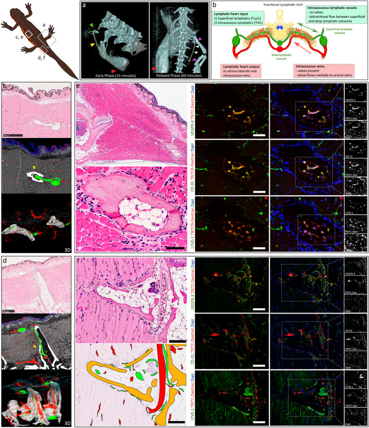
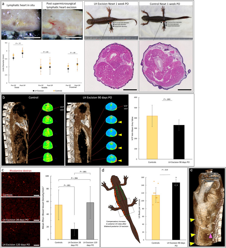
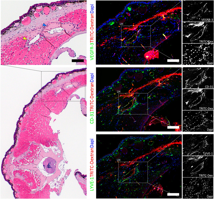
Regeneration competent vertebrates such as newts and salamanders possess a weakened adaptive immune system characterized by multiple connections between the lymphatic system and the blood vascular system called lymphatic hearts. The role of lymphatic vasculature and these lymphaticovenous connections in regeneration is unknown. We used in-vivo near-infrared lymphangiography, ultra-high frequency ultrasonography, micro-CT lymphangiography, and histological serial section 3-dimentional computer reconstruction to evaluate the lymphatic territories of Cynops pyrrhogaster. We used our model and supermicrosurgery to show that lymphatic hearts are not essential for lymphatic circulation and limb regeneration. Instead, newts possess a novel intraosseous network of lymphatics inside the bone expressing VEGFR-3, LYVE-1 and CD-31. However, we were unable to show Prox-1 expression by these vessels. We demonstrate that adult newt bone marrow functions as both a lymphatic drainage organ and fat reservoir. This study reveals the fundamental anatomical differences between the immune system of urodeles and mammals and provides a model for investigating lymphatics and regeneration.


Conflict of interest statement

The authors declare no competing interests.

Figures

Figure 1
Figure 1
Lymphatic territories mapped using in-vivo indocyanine green near infrared fluoroscopic lymphangiography. (a) Diagram showing the lymphatic flow territories of the newt forelimb (blue), hindlimb (yellow) and tail (green) mapped using indocyanine green (ICG) near infra-red fluoroscopy (NIRF). The trunci lymphatici longitudinales lateralis (TLyLL) (white arrowheads) run along the body interconnecting all the lymphatic hearts (LH) (green circles). (b) Forelimb lymphatics (blue arrowheads) run anteriorly to a dense axilla network and primarily drained into LH2 and 3 (green circles) then LH1, 3 and 5. Collecting lymphatics from the axilla crossed the mid-line on the ventral surface to join the collectors of the head region and the contralateral forelimb. The ICG injection sites are shown with red arrowheads. (c) Hindlimb lymphatics (yellow arrowheads) run posteriorly and flowed primarily to LH11 and LH12 (green circles) then to LH8, 9, 10, 13, 14 and 15. The hindlimb lymphatics connected to a dense network around the cloaca. Lymphatics on the right side (d) and left side (e) of the tail (green arrowheads) were drained by respective ipsilateral lymphatic vessels with the main collectors on the left and right running a slightly different course anteriorly and proceeded to form the TLyLL on each side. Lymphatic fluid from the tail drained into LH16 (green circle) and later LH15 through the TLyLL. (f) Diagram showing the lymphatic flow territories of the frog forelimb (blue) and hindlimb (magenta) mapped using ICG NIRF. The large subcutaneous lymphatic sacs (yellow) connecting the limb lymphatics and the LH (green circles). (g) The frog hindlimb was drained mainly by dorsal collectors (magenta arrowheads) to the posterior LHs (green circles) and some fluid flowed to the large subcutaneous lymphatic sacs (yellow outline). Forelimb lymphatics flowed rapidly into the lymphatic sacs obscuring the deeper lymphatic vasculature before flowing to the posterior LHs (blue arrowheads). The ICG injection sites are shown with red arrowheads. No subcutaneous lymphatic sacs were seen in newts.
Figure 2
Figure 2
Ultra-high frequency ultrasound-based calculation of the newt lymphatic heart cardiac output. (a) The lymphatic heart (LH) structure consists of a single muscular chamber receiving lymphatic fluid from the lymphatic collectors and pumping this fluid into the vena lateralis vein. The LH has one input valve and one output valve that prevent backflow of blood. Computer 3D volume reconstruction showed the newt lymphatic heart shape conformed best to an ellipsoid-like shape with a slightly pointed output end and flattened input end, therefore, the ejection fraction (EF) was calculated using an ellipsoid mathematical model based on 2D ultra-high frequency ultrasound measurement of the LH end diastolic volume (EDV) (b) and end systolic volume (c), and the LH rate. The mean EF was 60.40% (SD 12.94), EDV 0.026 mm3 (SD 0.008) and the mean lymphatic heart rate was 117.04 bpm (SD 23.12), giving a LH cardiac output (LHO) of 1.838 mm3/min for each LH and a combined total of 58.816 mm3/min for all 16 pairs.
Figure 3
Figure 3
Newt microcirculation and the intraosseous bone marrow lymphatic network. (a) Micro-CT Lymphangiography showed movement of contrast injected in the left hindlimb (red arrowhead) flowed through collectors (yellow arrowheads) to the lymphatic hearts (LH) (green arrowhead) and into the vertebral bone to the contralateral collectors (blue arrowhead), contralateral LHs (magenta arrowheads) and the trunci lymphatici longitudinales lateralis (TLyLL) (white arrowheads). (b) The functional lymphatic unit repeated throughout the trunk of the newt. The LH input from the superficial lymphatic network via the TLyLL and the transverse vertebral intraosseous lymphatics (TvIL) connecting the deep lymphatic network to the superficial lymphatic network and LHs on both sides of the body. (c) and (d) Serial section computer 3D volume reconstruction showing the lymphatic vessels (green) and blood vessels (red). The TvIL (yellow arrowheads) are shown flowing from the bone to the LHs. The anterior LHs of the trunk (c) were more closely attached to the adjacent rib bone while in the tail (d) they were separated from the bones by skeletal muscle. (e) and (f) Immunohistochemistry showing intraosseous lymphatic vessels and blood vessels in the bone marrow of the transverse process of an abdominal rib (Scale bars = 100 μm) (e) and the tail vertebra (Scale bars = 200 μm) (f). Lymphatic endothelial cells were identified by high rhodamine-dextran uptake and VEGFR-3 + , LYVE-1 + , CD-31 + staining while blood vessel endothelial cells were CD-31 + , LYVE-1 ± , VEGFR-3- and low rhodamine-dextran uptake.
Figure 4
Figure 4
Ultra-structure of the intraosseous lymphatics and veins and the lymphatic heart. (a) Resin embedded toluidine blue stained scanning section of a newt abdominal rib showing the transverse vertebral intraosseous lymphatics (TvIL) (b) and the transverse vertebral intraosseous veins (TvIV) (c) inside the bone marrow, and the adjacent lymphatic heart (LH) (d). Transmission Electron Microscopy confirmed the TvIL ((b) Scale bar = 2 μm) and LH ((d) Scale bar = 20 μm) were lined by lymphatic endothelial cells (LEC) with the characteristic ultra-structural features of lymphatic endothelium including absence of pericytes, thin walls and highly attenuated LEC cytoplasm (green arrowheads (b)), with sparse tight junctions, few adherens junctions, occasional spaces between LECs (yellow arrowheads (b and d')) and overlapping leaf-like endothelial cell junctions (blue arrowhead (d')). The specialized LH muscle had characteristic features of both cardiac and skeletal muscle (white arrowhead (d')). In contrast, the TvIV ((c) Scale bar = 5 μm) were lined by blood endothelial cells (BEC) had thicker walls, surrounded by pericytes (magenta arrowheads (c)) with several tight junctions and adherens junctions (red arrowheads (c)) and had multiple cytoplasm vesicles. Red blood cells were also seen in blood vessels.
Figure 5
Figure 5
Physiological changes in lymphatic circulation following lymphatic heart excision. (a) Lymphatic hearts (LH) excision in newts did not cause edema, histological changes, nor death (Scale bars = 1 mm). There was no significant difference in the 1-week post operative (PO) limb diameters (proximal t(9) = 0.729, p = 0.485, middle t(9) = − 0.504, p = 0.627, distal t(9) = 0.297, p = 0.773) between LH excision newts and controls with good inter-rater reliability ICC = 0.893, 95% CI (0.825, 0.934). (b) Micro-CT lymphangiography showed reduced lymphatic contrast in the blood circulation in LH excision newts at 90 days PO, with Trunci lymphatici longitudinales parabdominales (TLyLPab) collateral flow seen (yellow arrowheads). Vena cava posterior (VCP) analysis showed LH excision newts had a significantly lower mean grey value (t(34) = − 3.156, p = 0.003) than controls. (c) Rhodamine-dextran fluorescence lymphangiography analysis of lymphatics to vein flow (Scale bars = 100 μm) also showed a significant decrease in the density of blood vessels receiving lymphatic fluid (t(188) = 13.057, p < 0.001) at 28 days post excision of all the LHs but this returned to normal at 120 days PO (t(161) = − 0.854, p = 0.394). (d) Ultra-high frequency ultrasonography showed a significant increase (t(12) = − 2.707, p = 0.019) in the LH rate in newts that had bilateral posterior LH excision. (e) Collateral flow maintained lymphatic circulation after excision of LHs. TLyLPab collateral is shown (yellow arrowheads) with connections to the TvIL (magenta arrowhead). These results confirmed that LH excision successfully terminated peripheral lymphatic to vein flow for 90 days following which normal lymphatic to vein flow was restored by 120 days PO. Data represent mean ± standard deviation (error bars).
Figure 6
Figure 6
Morphological stages of lymphatic heart regeneration following complete excision. (a) Normal lymphatic heart (LH) (magenta arrowheads) with its supplying nerve (blue arrowheads), the trunci lymphatici longitudinales lateralis (TLyLL) (black arrowheads) and venous lateralis (VL) (red arrowheads). LH function was confirmed by collection of rhodamine-dextran (Scale bars = 100 μm). (b) The tissue defect immediately after LH excision. A segment of the TLyLL, VL and the supplying nerve, all of which are intimately associated with the LH were also resected. (c) Stage 1 Post Operative (PO) Day 5–10: Clot formation was followed by accumulation of a soft, fragile tissue mass beneath the wound epidermis. (d) Stage 2 PO Day 28–40: Angiogenesis of several small blood vessels interconnecting the cut ends of the VL and the other major blood vessels. No lymphangiogenesis seen at the injury site. Histology showed regenerating muscle and fibroblasts but no functional lymphatics with no rhodamine-dextran collection. (e) Stage 3 PO Day 50–60: Maturation of blood vessels restoring their original size and structure and restoration of VL continuity. Small lymphatic vessels were observed sprouting from the edges of the wound towards the injury zone. (f) Stage 4 PO Day 80–100: Lymphatic vessel maturation with active flow and collection of dye observed as a cystic mass in the region of the original LH, but no pulsations. (g) Stage 5 PO Day 100–130: Complete LH regeneration with pulsation. The regenerated LHs had fully functional input and output valves and uptake of rhodamine-dextran confirming they were fully functional.
Figure 7
Figure 7
Lymphatic endothelial cell marker expression in newly regenerated lymphatics. Histological sections showing the regenerated lymphatic heart (LH) (yellow arrowhead) and the adjacent rib bone (blue arrowhead) in the abdominal region 120 days post complete microsurgical excision. The newly regenerated LHs had identical shape, size, structure, position, and function as the original LH. In addition, the newly regenerated LH demonstrated strong expression of lymphatic endothelial cell markers VEGFR-3 and LYVE-1 and pan-endothelial marker CD-31. The uptake of rhodamine-dextran dye confirmed the lymphatics were fully functional (Scale bars = 200 μm).
Figure 8
Figure 8
Randomized control trial evaluating the result of lymphatic heart excision on the rate and morphology of limb regeneration. (a) The randomized control trial design: n = 62 newts were enrolled and separated into 4 blocks, each block was randomized into a study and control group by a blinded independent assistant. Follow-up was complete in 52 newts, 10 newts died immediately postoperative. (b) There was no statistically significant difference in the overall rate of regeneration between study and control groups (Kolmogorov–Smirnov Z = 0.662, p = 0.773). (ch) Subgroup analysis also showed no statistically significant difference in regeneration rates between study and control newts after unilateral LH excision (Kolmogorov–Smirnov Z = 0.730, p = 0 .660), regardless of the timing of amputation immediate amputation after LH excision (Kolmogorov–Smirnov Z = 0.617, p = 0.841) and amputation delayed 1-week post LH excision (Kolmogorov–Smirnov Z = 0.661, p = 0.774). Similarly, no significant difference between study and control newts was found after incremental bilateral LH excision (Kolmogorov–Smirnov Z = 0.778, p = 0.579) regardless of the timing of amputation immediate amputation (Kolmogorov–Smirnov Z = 1.284, p = 0.074) and amputation delayed 1-week PO (Kolmogorov–Smirnov Z = 1.193, p = 0.116). (i) Comparison of LH excision study newts from the different experimental batches (excluding controls) also showed no significant difference in the time taken to complete regeneration (Kruskal–Wallis H(3) = 0.1.229, p = 0.746). (j) Comparison of the morphological abnormalities in the regenerated limbs showed no statistically significant difference (p = 0.150) between study and control newts. Data represent mean ± standard deviation (error bars).

References

    1. Hansen KC, D’Alessandro A, Clement CC, Santambrogio L. Lymph formation, composition and circulation: A proteomics perspective. Int. Immunol. 2015;27:219–227. doi: 10.1093/intimm/dxv012. - DOI - PubMed
    1. Dzieciatkowska M, et al. Lymph is not a plasma ultrafiltrate: A proteomic analysis of injured patients. Shock. 2014;42:485–498. doi: 10.1097/SHK.0000000000000249. - DOI - PMC - PubMed
    1. Ulvmar MH, Mäkinen T. Heterogeneity in the lymphatic vascular system and its origin. Cardiovasc. Res. 2016;111:310–321. doi: 10.1093/cvr/cvw175. - DOI - PMC - PubMed
    1. Randolph GJ, Ivanov S, Zinselmeyer BH, Scallan JP. The lymphatic system: Integral roles in immunity. Annu. Rev. Immunol. 2017;35:31–52. doi: 10.1146/annurev-immunol-041015-055354. - DOI - PMC - PubMed
    1. Maruyama K, Imanaka-Yoshida K. The pathogenesis of cardiac fibrosis: A review of recent progress. Int. J. Mol. Sci. 2022;23:2617. doi: 10.3390/ijms23052617. - DOI - PMC - PubMed

Publication types