Review
. 2023 Aug;24(8):576-596.
doi: 10.1038/s41580-023-00604-z. Epub 2023 Apr 27.

Regulation of phospholipid distribution in the lipid bilayer by flippases and scramblases

Affiliations
Review

Regulation of phospholipid distribution in the lipid bilayer by flippases and scramblases

Takaharu Sakuragi et al. Nat Rev Mol Cell Biol. 2023 Aug.

Erratum in

Abstract

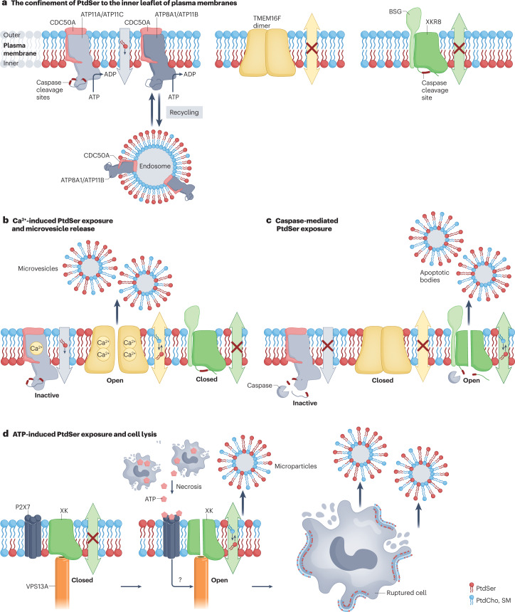
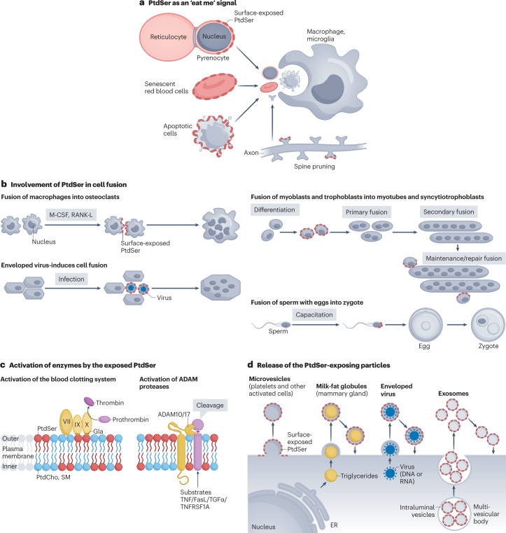
Cellular membranes function as permeability barriers that separate cells from the external environment or partition cells into distinct compartments. These membranes are lipid bilayers composed of glycerophospholipids, sphingolipids and cholesterol, in which proteins are embedded. Glycerophospholipids and sphingolipids freely move laterally, whereas transverse movement between lipid bilayers is limited. Phospholipids are asymmetrically distributed between membrane leaflets but change their location in biological processes, serving as signalling molecules or enzyme activators. Designated proteins - flippases and scramblases - mediate this lipid movement between the bilayers. Flippases mediate the confined localization of specific phospholipids (phosphatidylserine (PtdSer) and phosphatidylethanolamine) to the cytoplasmic leaflet. Scramblases randomly scramble phospholipids between leaflets and facilitate the exposure of PtdSer on the cell surface, which serves as an important signalling molecule and as an 'eat me' signal for phagocytes. Defects in flippases and scramblases cause various human diseases. We herein review the recent research on the structure of flippases and scramblases and their physiological roles. Although still poorly understood, we address the mechanisms by which they translocate phospholipids between lipid bilayers and how defects cause human diseases.


Conflict of interest statement

The authors declare no competing interests.

Figures

Fig. 1
Fig. 1. Flippases and scramblases that regulate the phospholipid distribution in the lipid bilayer.
a, Phospholipids. Glycerophospholipids are phosphatic acid (PA) derivatives composed of glycerol attached to two fatty acyl chains (diacylglycerol; DAG) and a phosphate. Serine, choline, or ethanolamine is conjugated with the phosphate of PA to produce phosphatidylserine (PtdSer), phosphatidylcholine (PtdCho) or phosphatidylethanolamine (PtdEtn). Sphingomyelin (SM) is a ceramide to which phosphocholine is attached. b, Flippases and scramblase. Under normal conditions, ATP-driven flippases translocate PtdSer and PtdEtn from the outer or lumen side to the inner leaflets of the lipid bilayer to maintain their confined localization to the inner leaflet. Scramblases non-specifically translocate phospholipids to disrupt the asymmetric distribution of phospholipids and to expose PtdSer on the outer or lumen side of cell membranes. c, Transfer of PtdSer from the endoplasmic reticulum (ER) to plasma membranes. PtdSer synthesized by PtdSer synthase at the ER is transferred to plasma membranes via a reciprocal exchange with PtdIns(4)P by a lipid transfer protein of the Oxysterol-binding protein (OSBP)-related protein (ORP) family. PSS, phosphatidyl serine synthase.
Fig. 2
Fig. 2. Asymmetrical distribution of PtdSer and its breakdown.
a, Type IV P-type ATPase (P4-ATPase)-mediated confinement of phosphatidylserine (PtdSer) to the inner leaflet of the plasma membrane. ATP11A and ATP11C are complexed with CDC50A at the plasma membrane and serve as flippases to specifically translocate PtdSer from the outer leaflet to the inner leaflet in growing cells. ATP8A1 and ATP11B seem to recycle between the endosomes and plasma membranes and maintain the asymmetrical distribution of PtdSer at the plasma membrane. b, Ca2+-induced PtdSer exposure and microvesicle release. When platelets, osteoblasts and other cells are activated, the intracellular Ca2+ concentration increases. Binding of Ca2+ to ATP11A and ATP11C inhibits their flippase activity while activating TMEM16F, which causes temporal PtdSer exposure on the cell surface and the release of microvesicles. These microvesicles also expose PtdSer, which conveys further functions (see also Fig. 4c). c, When cells undergo apoptosis, caspase 3 is activated and cleaves the carboxy-terminal part of XKR8 to trigger its scrambling activity. At the same time, caspase 3 cleaves ATP11A and ATP11C in the middle of the molecule to inactivate them, allowing the cells to expose PtdSer irreversibly. This process is accompanied by the release of apoptotic bodies, which also expose PtdSer and are engulfed by phagocytes. d, ATP-induced PtdSer exposure and cell lysis. XK, a member of the XK-related (XKR) family, is complexed with VPS13A lipid transporter via its β-hairpin in the centre of the molecule. ATP released from necrotic cells binds the P2X7 homotrimer, and an unidentified signal (question mark) from the ATP-engaged P2X7 receptor activates the XK–VPS13A complex to scramble phospholipids in the plasma membrane. This causes the PtdSer exposure on the cell surface, followed by cell rupture. PtdCho, phosphatidylcholine; SM, sphingomyelin; TMEM16, transmembrane protein 16.
Fig. 3
Fig. 3. Models for lipid transport.
a, Credit card model for lipid transport. The hydrophilic head group of the phospholipid (corresponding to the magnetic strip on the credit card) is protected by the hydrophilic groove of the transporter (track of the card reader) during lipid transport. Image reproduced from Pomorski et al., ref. . b, Structure of ATP11C–CDC50A and a model for lipid flipping. Tertiary structure of human ATP11C and CDC50A complex in a phosphatidylserine (PtdSer) occluded E2-Pi state [PDB:7BSV] shown on the left. P4-ATPases are coloured grey with selected helices (α1, α2, α4 and α6) in blue, whereas CDC50A is red. Actuator domain (A) which dephosphorylates the phosphorylated P site shown in yellow. PtdSer is coloured green–red–white. Right panel shows the proposed lipid transport cycle. In an outward open state (E2P), in which the phosphorylation (P) site is phosphorylated (P in yellow circle), a phospholipid binds to the external open cavity. In the phospholipid occluded state (E2-Pi), the phosphate is detached from the P site (Pi in a yellow circle), and helices α1 and α2 move to cover the ectoplasmic side of the cavity (red arrow). In the inward open state (E2), the phosphate (Pi in yellow circle) is released from the molecule (blue arrow), which is coupled by the movement of the A domain to the periphery (black arrow). This changes the arrangement of helices α1 and α2 (green arrow), and the cavity is now accessible to the cytosolic leaflet releasing the phospholipid. c, Structure of transmembrane protein 16 (TMEM16) and model for lipid scrambling. The Ca2+-bound open state structure of Nectria haematococca (nh)TMEM16 [PDB:4WIS] is viewed from the front and side (left). Each protomer of TMEM16 is coloured grey or dark grey. Selected helices (α3, α4, α5 and α6) are coloured blue and labelled. On the right, the activation and scrambling, viewed from the side, are schematically depicted. In a Ca2+-free closed state, the cavity is shielded from the membrane with helices α4 and α6 tightly interacting. The binding of Ca2+ triggers the conformational change to the open state; helices α4 and α6 are separated, and the cavity is accessible to the phospholipid head group. d, Structure of XK-related 8 (XKR8) and a model for lipid scrambling. The tertiary structure of the human XKR8–Basigin complex [PDB:7DCE] is shown with phosphatidylcholine (PtdCho) bound to the hydrophobic cleft. Selected helices (α1, α2, α4 and α11) are coloured blue and labelled. A model for the caspase-activated scrambling mechanism is shown on the right. A phospholipid is recruited from the outer leaflet to the hydrophobic cleft, even in a closed state. When cells undergo apoptosis, caspase 3 cleaves helix 11, which likely induces the movement of helix 2 (red arrows) to expose the path for phospholipid transport.
Fig. 4
Fig. 4. Biological role of PtdSer.
a, Phosphatidylserine (PtdSer) as an ‘eat me’ signal. When cells undergo apoptosis, they expose PtdSer (red ellipse) on the surface. They are then dismantled into apoptotic bodies with PtdSer on their surface. These bodies are recognized by phagocytes via TAM receptors and swiftly eliminated to ensure the removal of cellular debris and immunogenic reactions. At the final stage of definitive erythropoiesis, erythroblasts divide into reticulocytes and pyrenocytes. Soon after the separation from reticulocytes, the pyrenocytes expose PtdSer and are engulfed by macrophages. Human red blood cells have a lifetime of about 120 days. The senescent red blood cells expose PtdSer for engulfment by macrophages. Similar mechanisms operate in neurons, whose membranes undergo regular, experience-driven pruning to remove unconnected or damaged dendritic spines (the primary location for excitatory synapses). These dendritic spines expose PtdSer, which is recognized by microglia for their removal. b, Involvement of PtdSer in cell fusion. Fusion of macrophages into osteoclasts: when macrophages are treated with macrophage colony-stimulating factor (M-CSF) or receptor activator of nuclear factor-κB ligand (RANKL), they fuse into giant osteoclasts, during which PtdSer (red ellipse) is transiently exposed to the cell surface promoting fusion. Enveloped virus-mediated cell fusion: when an enveloped virus infects cells, they fuse in a PtdSer-dependent manner. Fusion of myoblasts and trophoblasts: myoblasts and trophoblasts differentiate and fuse to form myotubes and syncytiotrophoblasts, respectively, which involves exposed PtdSer. The fusion process is also important for further growth and membrane repair in myotubes and syncytiotrophoblasts. Fusion of sperm with eggs into zygotes: before fertilization, sperm are capacitated in the female reproductive tract, which is accompanied by the PtdSer exposure on the head region. The blocking PtdSer inhibits the fusion process. Many molecules have been proposed for this process. However, very little is known about how cell fusion proceeds. c, Activation of enzymes by the exposed PtdSer. Activation of the blood clotting system: during blood clot formation, platelets are activated to expose PtdSer. This allows the binding and activation of factors VII, IX and X, which recognizes PtdSer via its ‘Gla’ domain. Factor X forms a complex with factor VII and factor IX and mediates the cleavage of prothrombin (factor II) into thrombin for blood clotting. Activation of a disintegrin and metalloproteinase (ADAM) proteases: membrane proteases, ADAM10 and ADAM17, are activated by the exposed PtdSer and work to cleave TNF, Fas ligand, TGFα, secretin, TNF receptor superfamily member 1A (TNFRSF1A) and others, releasing them into the microenvironment to allow paracrine signalling. d, Release of the PtdSer-exposing particles. When platelets and osteoblasts are activated, they expose PtdSer and produce microvesicles, which also expose PtdSer. The platelet-derived microvesicles are involved in propagating the blood clot cascade (see part c), whereas microvesicles released by osteoblasts (called matrix vesicles) carry concentrated phosphate and Ca2+ that form hydroxyapatite crystal — a process required for bone mineralization and supported by PtdSer, which has been proposed to act as a nucleator for the crystallization process. Mammary glands secrete milk fat globules (MFGs) — triglycerides covered by plasma membranes exposing PtdSer. Enveloped viruses are nucleic acids surrounded by the PtdSer-exposing plasma membrane. Exosomes derived from the multivesicular bodies are secreted from various cells and expose PtdSer. MFGs, enveloped viruses and exosomes may rebind to the cell surface in a PtdSer-dependent manner, supporting their uptake. ER, endoplasmic reticulum; PtdCho, phosphatidylcholine; SM, sphingomyelin.

References

    1. Vance JE. Phospholipid synthesis and transport in mammalian cells. Traffic. 2015;16:1–18. doi: 10.1111/tra.12230. - DOI - PubMed
    1. Yang Y, Lee M, Fairn GD. Phospholipid subcellular localization and dynamics. J. Biol. Chem. 2018;293:6230–6240. doi: 10.1074/jbc.R117.000582. - DOI - PMC - PubMed
    1. Bretscher MS. Asymmetrical lipid bilayer structure for biological membranes. Nat. N. Biol. 1972;236:11–12. doi: 10.1038/newbio236011a0. - DOI - PubMed
    1. Tsuji T, et al. Predominant localization of phosphatidylserine at the cytoplasmic leaflet of the ER, and its TMEM16K-dependent redistribution. Proc. Natl Acad. Sci. USA. 2019;116:13368–13373. doi: 10.1073/pnas.1822025116. - DOI - PMC - PubMed
    1. Murate M, et al. Transbilayer distribution of lipids at nano scale. J. Cell Sci. 2015;128:1627–1638. - PubMed

Publication types

MeSH terms