. 2023 Mar 27;10(4):423.
doi: 10.3390/bioengineering10040423.

Proteomic Profiling of Fallopian Tube-Derived Extracellular Vesicles Using a Microfluidic Tissue-on-Chip System

Affiliations

Proteomic Profiling of Fallopian Tube-Derived Extracellular Vesicles Using a Microfluidic Tissue-on-Chip System

Didi Zha et al. Bioengineering (Basel). .

Abstract

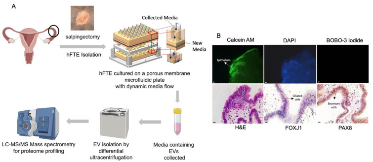
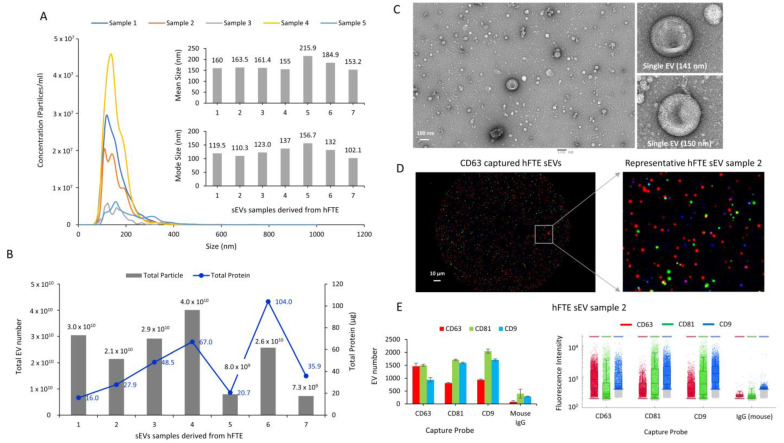
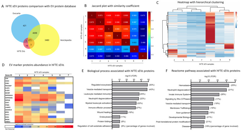
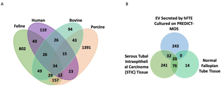
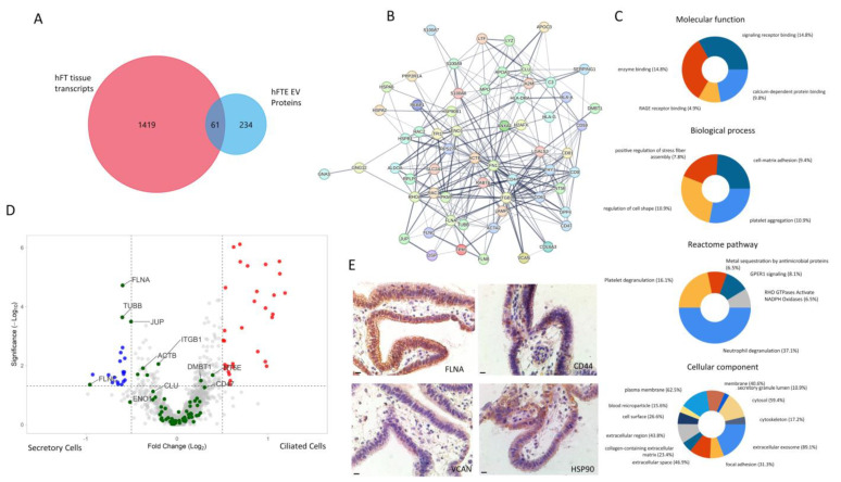
The human fallopian tube epithelium (hFTE) is the site of fertilization, early embryo development, and the origin of most high-grade serous ovarian cancers (HGSOCs). Little is known about the content and functions of hFTE-derived small extracellular vesicles (sEVs) due to the limitations of biomaterials and proper culture methods. We have established a microfluidic platform to culture hFTE for EV collection with adequate yield for mass spectrometry-based proteomic profiling, and reported 295 common hFTE sEV proteins for the first time. These proteins are associated with exocytosis, neutrophil degranulation, and wound healing, and some are crucial for fertilization processes. In addition, by correlating sEV protein profiles with hFTE tissue transcripts characterized using GeoMx® Cancer Transcriptome Atlas, spatial transcriptomics analysis revealed cell-type-specific transcripts of hFTE that encode sEVs proteins, among which, FLNA, TUBB, JUP, and FLNC were differentially expressed in secretory cells, the precursor cells for HGSOC. Our study provides insights into the establishment of the baseline proteomic profile of sEVs derived from hFTE tissue, and its correlation with hFTE lineage-specific transcripts, which can be used to evaluate whether the fallopian tube shifts its sEV cargo during ovarian cancer carcinogenesis and the role of sEV proteins in fallopian tube reproductive functions.

Keywords: digital spatial imaging; extracellular vesicles; fallopian tube; microfluidic culture; proteomics.


Conflict of interest statement

A.K.G. is a co-founder of Sinochips Diagnostics, serves as a scientific advisory board member to Biovica, Clara Biotech, and Sinochips Diagnostics, and receives research funding from Predicine, Inc. and VITRAC Therapeutics. The other authors report no conflicts of interest.

Figures

Figure 1
Figure 1
The fallopian tube explants were cultured in a microfluidic platform for sEV collection. (A) The workflow of hFTE-derived EV proteomics characterization. (B) The live/dead staining of hFTE epithelium cultured in the PREDICT-MOS for six days. The green fluorescence (calcein) indicates live cells, and the red fluorescence indicates dead cells, labeled by BOBO-3 iodide. Immunohistochemistry staining of FOXJ1 and PAX8 confirmed that the hFTE maintained the two major epithelial cell populations, ciliated and secretory cells, after six-day dynamic culture on the PREDICT-MOS. Scale bar = 10 µm.
Figure 2
Figure 2
The physical and molecular characterization of sEVs derived from hFTE cultured in the PREDICT-MOS. (A) The size profile of sEVs was quantified using nanoparticle-tracking analysis (NTA). NTA graphs of five representative samples are shown. Inset shows the mean size and mode size of sEVs from seven patient samples. (B) The total sEV number and total protein content in sEVs quantified via NTA and the Bradford assay, respectively. (C) The transmission electron microscopy (TEM) image of sEVs shows cup-shaped morphological properties. Inset shows the micrograph of a single sEVs (Scale bar: 100 nm). (D) The image of sEVs captured by the CD63 capture probe in ExoView chip, and detected by CD63 (red), CD81 (green), and CD9 (blue) antibodies (Scale bar: 10 µm). (E) The molecular characterization of sEVs quantifying CD63/CD81/CD9-positive sEVs using the ExoView platform. The graph shows the particle number and fluorescent intensity of sEVs captured by the CD63/CD81/CD9 probe, and detected by CD63/CD81/CD9 fluorescent antibodies in representative sEV samples.
Figure 3
Figure 3
The proteomics characterization of sEVs derived from hFTE cultured in the PREDICT −MOS. (A) The comparison of 295 common proteins identified in at least five of seven hFTE sEV samples, with established EV protein databases, Vesiclepedia and Exocarta, showing 275 common proteins and 19 unique proteins. (B) The plot referring to the Jaccard index (or similarity coefficient), assessing the similarity in terms of the number of expressed proteins among seven samples based on 295 proteins. A Jaccard index of 1 (diagonal) indicates that the same proteins are expressed (100% similarity). A value of 0 for the Jaccard index would indicate no similarity corresponding to completely different sets of expressed proteins (no overlap or 0% similarity). (C) A heatmap demonstrating the protein abundance of 295 proteins among seven hFTE sEV samples. Rows are centered, and unit-variance scaling is applied to rows. Both rows and columns are clustered by Pearson’s correlation as a method, and Ward as a distance. (D) A heatmap showing the expression of proteins most frequently identified in EVs, based upon Vesiclepedia and Exocarta databases among seven hFTE sEVs samples. (E,F) Gene-enrichment analysis using GO biological processes and REACTOME pathway analysis, showing the biological processes and pathways associated with 295 hFTE sEV proteins. Inset in the figure represents the percentage of the genes responsible for a particular biological process.
Figure 4
Figure 4
hFTE sEV protein content comparison across species and FT lesions. (A) Venn diagram showing the number of sEV proteins identified exclusively in human fallopian tube epithelium (hFTE), bovine, porcine, cat oviduct, or in common [3,33,34]. (B) Venn diagram showing shared proteins identified in STIC tissue [35] and proteins found in hFTE-derived sEVs cultured in the microfluidic device, PREDICT-MOS.
Figure 5
Figure 5
Correlation of fallopian tube tissue transcriptomics profile with a hFTE sEV proteomics profile. (A) Comparison between hFTE sEV proteins with hFT tissue transcripts (CTA database, GeoMx) of ciliated and secretory cells identified by GeoMx digital spatial imaging showing 61 common gene IDs. (B) Protein−protein interaction network of the 61 common gene IDs via STRING. (C) Gene enrichment analysis of 61 common genes showing molecular functions, biological processes, Reactome pathways, and cellular components associated with 61 common genes, using the FUNRICH software. The percentage in the pie chart description represents the percentage of genes out of 61 common genes responsible for particular events. (D) The differential expression of genes in ciliated vs. secretory cells in fallopian tube tissue. The green dots represent 61 common gene IDs between the hFTE transcriptome and hFTE sEV proteomics. Four (FLNA, TUBB, JUP, and FLNC) out of 61 genes were significantly upregulated in secretory cells. Blue and red dots are differentially expressed genes in secretory and ciliated cells, respectively. (E) IHC staining of representative proteins of 61 common genes expressed in fallopian tube tissue and sEVs. Scale bar = 10 µm.

References

    1. Li S., Winuthayanon W. Oviduct: Roles in Fertilization and Early Embryo Development. J. Endocrinol. 2017;232:R1–R26. doi: 10.1530/JOE-16-0302. - DOI - PubMed
    1. Almiñana C., Heath P.R., Wilkinson S., Sanchez-Osorio J., Cuello C., Parrilla I., Gil M.A., Vazquez J.L., Vazquez J.M., Roca J., et al. Early Developing Pig Embryos Mediate Their Own Environment in the Maternal Tract. PLoS ONE. 2012;7:e33625. doi: 10.1371/journal.pone.0033625. - DOI - PMC - PubMed
    1. Almiñana C., Corbin E., Tsikis G., Alcântara-Neto A.S., Labas V., Reynaud K., Galio L., Uzbekov R., Garanina A.S., Druart X., et al. Oviduct Extracellular Vesicles Protein Content and Their Role during Oviduct–Embryo Cross-Talk. Reproduction. 2017;154:253–268. doi: 10.1530/REP-17-0054. - DOI - PubMed
    1. Winuthayanon W., Bernhardt M.L., Padilla-Banks E., Myers P.H., Edin M.L., Lih F.B., Hewitt S.C., Korach K.S., Williams C.J. Oviductal Estrogen Receptor α Signaling Prevents Protease-Mediated Embryo Death. eLife. 2015;4:e10453. doi: 10.7554/eLife.10453. - DOI - PMC - PubMed
    1. Georgiou A.S., Sostaric E., Wong C.H., Snijders A.P.L., Wright P.C., Moore H.D., Fazeli A. Gametes Alter the Oviductal Secretory Proteome. Mol. Cell. Proteom. 2005;4:1785–1796. doi: 10.1074/mcp.M500119-MCP200. - DOI - PubMed