Review
. 2023 Mar 28;12(4):508.
doi: 10.3390/biology12040508.

Inputs and Outputs of the Mammalian Circadian Clock

Affiliations
Review

Inputs and Outputs of the Mammalian Circadian Clock

Ashley N Starnes et al. Biology (Basel). .

Abstract

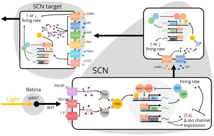
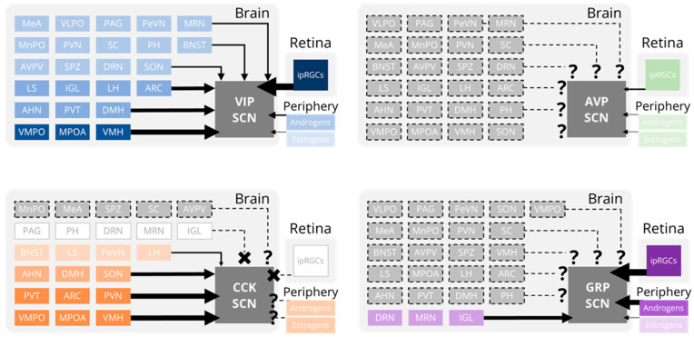
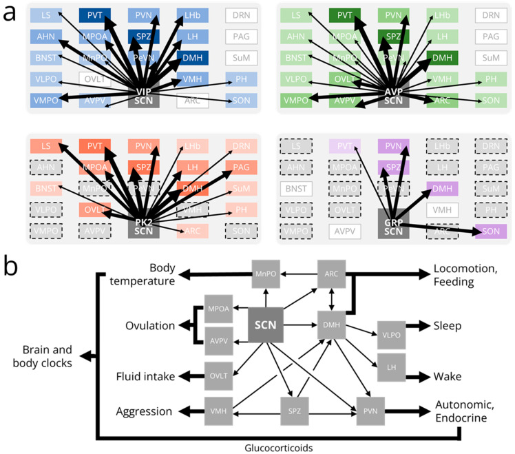
Circadian rhythms in mammals are coordinated by the central circadian pacemaker, the suprachiasmatic nucleus (SCN). Light and other environmental inputs change the timing of the SCN neural network oscillator, which, in turn, sends output signals that entrain daily behavioral and physiological rhythms. While much is known about the molecular, neuronal, and network properties of the SCN itself, the circuits linking the outside world to the SCN and the SCN to rhythmic outputs are understudied. In this article, we review our current understanding of the synaptic and non-synaptic inputs onto and outputs from the SCN. We propose that a more complete description of SCN connectivity is needed to better explain how rhythms in nearly all behaviors and physiological processes are generated and to determine how, mechanistically, these rhythms are disrupted by disease or lifestyle.

Keywords: circadian; circuits; suprachiasmatic.


Conflict of interest statement

The authors declare no conflict of interest.

Figures

Figure 1
Figure 1
Photoentrainment of the SCN and SCN target neurons. Light activates intrinsically-photosensitive retinal ganglion cells (ipRGCs) which project through the retinohypothalamic tract (RHT) to release glutamate (GLU) and pituitary adenylate cyclase-activating peptide (PACAP) onto retinorecipient suprachiasmatic nucleus (SCN) neurons [9,10]. The activation of glutamate receptors increases intracellular calcium ([Ca]i) directly through N-methyl D-aspartate (NMDA) receptors and indirectly through voltage-gated calcium channels (VGCCs) [11]. The activation of PAC1 receptors initiates a G protein cascade that increases intracellular cyclic AMP (cAMP). cAMP and [Ca]i activate protein kinase A (PKA) and calcium-dependent protein kinases (CAMK) to phosphorylate cAMP response element (CRE)-binding protein (CREB) [9,12]. Phosphorylated CREB promotes the transcription of the Period genes within the molecular circadian clock which increases the transcription of clock-controlled genes (CCGs) [13]. The translation of CCGs increases neuronal firing rate by directly increasing [Ca]i or by altering membrane ion channel expression [14,15,16]. Increased firing rate in retinorecipient SCN neurons increases the release of gamma-aminobutyric acid (GABA) and vasoactive intestinal peptide (VIP) onto other neurons in the SCN neural network [17,18]. This changes neuronal firing rate and transcription of core clock genes either directly through the activation of ionotropic GABAA receptors or indirectly through metabotropic GABAB receptors and the VIP receptor VPAC2 [19,20,21]. SCN output neurons subsequently release several output factors including GABA, VIP, arginine vasopressin (AVP), prokineticin 2 (PK2), transforming growth factor-alpha (TGFα), and cardiotrophin-like cytokine (CLC) onto SCN target neurons [22,23,24,25,26,27]. These SCN output signals ultimately entrain SCN target neurons by changing their firing rates and core clock gene transcriptional activity.
Figure 2
Figure 2
Inputs to the SCN. Schematics depicting the relative strengths of synaptic and hormonal inputs onto vasoactive intestinal peptide (VIP, blue), arginine vasopressin (AVP, green), cholecystokinin (CCK, orange), and gastrin-releasing peptide (GRP, purple) producing suprachiasmatic nucleus (SCN) neurons. Darker colors and thicker arrows represent stronger inputs from a given brain region or hormone. Gray boxes with no dashed outlines indicate brain regions with no inputs to an SCN subpopulation. Gray boxes with dashed outlines indicate brain regions whose (absence of) projections to an SCN subpopulation have not been experimentally determined. AHN, anterior hypothalamic nucleus; ARC, arcuate nucleus; AVPV, anteroventral periventricular nucleus; BNST, bed nucleus of the stria terminalis; DMH, dorsomedial hypothalamic nucleus; DRN, dorsal raphe nucleus; IGL, intergeniculate leaflet; ipRGCs, intrinsically-photosensitive retinal ganglion cells; LH, lateral hypothalamus; LS, lateral septum; MPOA, medial preoptic area; MRN, median raphe nucleus; MeA, medial amygdala; MnPO, median preoptic area; PAG, periaqueductal gray; PH, posterior hypothalamus; PVN, paraventricular nucleus of the hypothalamus; PVT, paraventricular nucleus of the thalamus; PeVN, periventricular nucleus; SC, superior colliculus; SON, supraoptic nucleus; SPZ, subparaventricular zone; VLPO, ventrolateral preoptic area; VMH, ventromedial hypothalamus; VMPO, ventromedial preoptic nucleus.
Figure 3
Figure 3
Outputs from the SCN. (a) Schematics depicting the relative strengths of synaptic outputs from vasoactive intestinal peptide (VIP, blue), arginine vasopressin (AVP, green), prokineticin 2 (PK2, red), and gastrin-releasing peptide (GRP, purple) producing suprachiasmatic nucleus (SCN) neurons. Boxes are roughly organized such that the brain region in the top left is the most rostral and most dorsal. Darker colors and thicker arrows represent stronger inputs onto a given brain region. Gray boxes with no dashed outlines indicate brain regions with no inputs from an SCN subpopulation. Gray boxes with dashed outlines indicate brain regions whose (absence of) inputs from an SCN subpopulation have not been experimentally determined. Note that the depicted GRP efferent projections have not been identified using modern genetic tools. (b) Schematic depicting output pathways from the SCN to several rhythmic behaviors and physiological processes. Glucocorticoid release and body temperature rhythms allow the SCN to synchronize distant brain and body clocks. AHN, anterior hypothalamic nucleus; ARC, arcuate nucleus; AVPV, anteroventral periventricular nucleus; BNST, bed nucleus of the stria terminalis; DMH, dorsomedial hypothalamus; DRN, dorsal raphe nucleus; LH, lateral hypothalamus; LHb, lateral habenula; LS, lateral septum; MPOA, medial preoptic area; MnPO, median preoptic area; OVLT, organum vasculosum of the lamina terminalis; PAG, periaqueductal gray; PH, posterior hypothalamus; PVN, paraventricular nucleus of the hypothalamus; PVT, paraventricular nucleus of the thalamus; PeVN, periventricular nucleus; SON, supraoptic nucleus; SPZ, subparaventricular zone; SuM, supramammillary nucleus; VLPO, ventrolateral preoptic nucleus; VMH, ventromedial hypothalamus; VMPO; ventromedial preoptic nucleus.

References

    1. Silver R., Rainbow M. The Suprachiasmatic Nucleus and the Circadian Timekeeping System of the Body. In: Pfaff D.W., editor. Neuroscience in the 21st Century: From Basic to Clinical. Springer; New York, NY, USA: 2013. pp. 1847–1888.
    1. Hastings M.H., Maywood E.S., Brancaccio M. Generation of Circadian Rhythms in the Suprachiasmatic Nucleus. Nat. Rev. Neurosci. 2018;19:453–469. doi: 10.1038/s41583-018-0026-z. - DOI - PubMed
    1. Moore R.Y., Speh J.C. GABA Is the Principal Neurotransmitter of the Circadian System. Neurosci. Lett. 1993;150:112–116. doi: 10.1016/0304-3940(93)90120-A. - DOI - PubMed
    1. Van den Pol A.N. The Hypothalamic Suprachiasmatic Nucleus of Rat: Intrinsic Anatomy. J. Comp. Neurol. 1980;191:661–702. doi: 10.1002/cne.901910410. - DOI - PubMed
    1. Ono D., Honma K.-I., Honma S. Roles of Neuropeptides, VIP and AVP, in the Mammalian Central Circadian Clock. Front. Neurosci. 2021;15:650154. doi: 10.3389/fnins.2021.650154. - DOI - PMC - PubMed

LinkOut - more resources