. 2023 Apr 12;12(4):589.
doi: 10.3390/biology12040589.

Structure-Function of the Human WAC Protein in GABAergic Neurons: Towards an Understanding of Autosomal Dominant DeSanto-Shinawi Syndrome

Affiliations

Structure-Function of the Human WAC Protein in GABAergic Neurons: Towards an Understanding of Autosomal Dominant DeSanto-Shinawi Syndrome

Hannah C Rudolph et al. Biology (Basel). .

Abstract

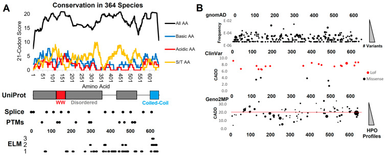
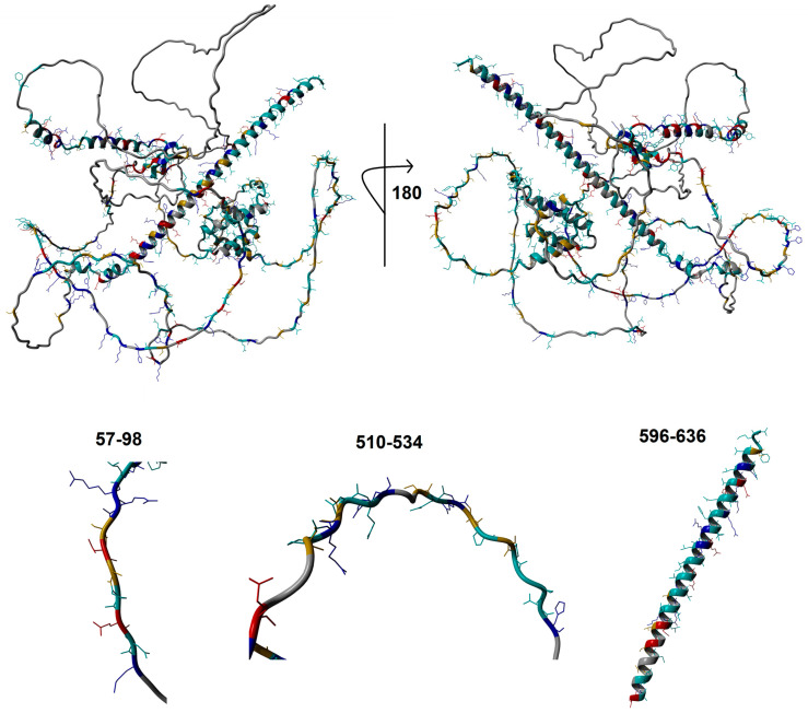
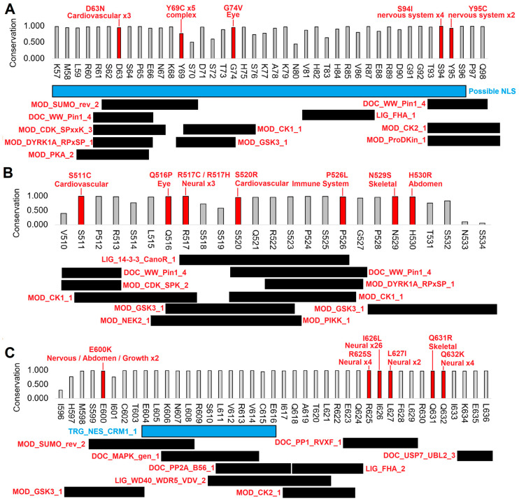
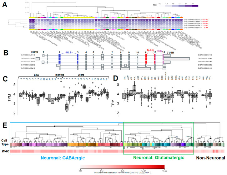
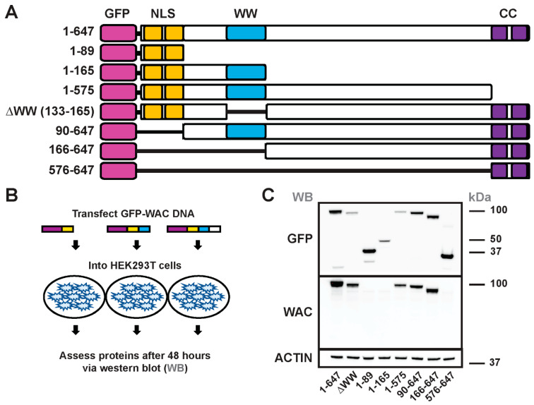
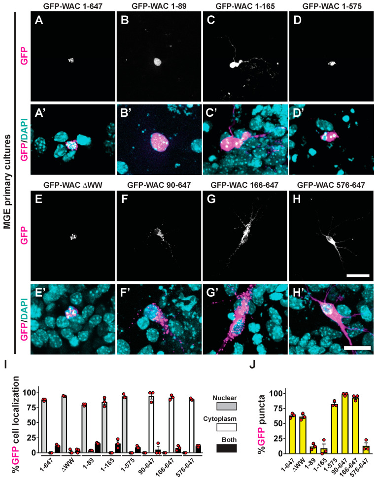
Dysfunction of the WW domain-containing adaptor with coiled-coil, WAC, gene underlies a rare autosomal dominant disorder, DeSanto-Shinawi syndrome (DESSH). DESSH is associated with facial dysmorphia, hypotonia, and cognitive alterations, including attention deficit hyperactivity disorder and autism. How the WAC protein localizes and functions in neural cells is critical to understanding its role during development. To understand the genotype-phenotype role of WAC, we developed a knowledgebase of WAC expression, evolution, human genomics, and structural/motif analysis combined with human protein domain deletions to assess how conserved domains guide cellular distribution. Then, we assessed localization in a cell type implicated in DESSH, cortical GABAergic neurons. WAC contains conserved charged amino acids, phosphorylation signals, and enriched nuclear motifs, suggesting a role in cellular signaling and gene transcription. Human DESSH variants are found within these regions. We also discovered and tested a nuclear localization domain that impacts the cellular distribution of the protein. These data provide new insights into the potential roles of this critical developmental gene, establishing a platform to assess further translational studies, including the screening of missense genetic variants in WAC. Moreover, these studies are essential for understanding the role of human WAC variants in more diverse neurological phenotypes, including autism spectrum disorder.

Keywords: WAC; cell biology; nuclear translocation; protein domain; protein sorting.


Conflict of interest statement

The authors declare no conflict of interest.

Figures

Figure 1
Figure 1
Genomic analysis of WAC. (A) Compiled genomics insights for WAC. On top is the conservation of 364 species of WAC protein. The conservation score of all amino acids (black) is based on the amino acid % of species conserved at each amino acid (0—no conservation, 1–100% conserved), placed on a 21 codon sliding window such that each site is added to 10 upstream and 10 downstream. The basic (blue, R/K/H), acidic (red, D/E), or S/T (orange) amino acid scores are calculated using only these amino acids for the same 21 codon window. Below is conservation of the amino acid locations (x-axis) for UniProt annotated domains/motifs, splice sites, posttranslational modifications (PTMs), and ELM annotated motifs (y-axis lists the number of annotations at each amino acid). (B) Extracted genomic variants from gnomAD (y-axis list allele frequency, bubble size is the number of unique variants), ClinVar (y-axis is the CADD functional score, color represents the type of variant), and Geno2MP variants (y-axis is the CADD score, bubble size the number of human phenotype profiles). Abbreviations—AA, amino acid; R, arginine; S, serine; T, threonine; LoF, loss-of-function; ELM, eukaryotic linear motif; HPO, human phenotype ontology.
Figure 2
Figure 2
Predicted WAC protein structure. An AlphaFold protein model of WAC showing conserved amino acids colored as blue: polar basic; red: polar acidic; orange: S/T; cyan: all other conserved amino acids. Shown below are zoomed in view of several conserved motifs within WAC that fall within intrinsically disordered regions.
Figure 3
Figure 3
Highly conserved motifs within WAC containing human variants connected to medical phenotypes. Shown for the N-terminal nuclear localization motif (A), central 14-3-3 interaction motif (B), and the C-terminal nuclear export motif region of the coiled-coil domain (C). The conservation is based on the 364 amino acid sequences of vertebrate WAC (0—no conservation, 1–100% conserved). Those amino acids in red have a known Geno2MP variant with listed phenotypes. Those with multiple individuals with overlapping phenotypes are represented with an x number of individuals. Below each region are ELM-predicted motifs.
Figure 4
Figure 4
Analysis of WAC expression in various tissues, the brain developmental times, and within single-cell types of human brain. (A) GTEx expression of WAC isoforms in various human tissues. The darker blue color represents higher expression. Tissues are ranked in expression. In red next to each isoform number is the amino acid size of the protein. (B) Annotated exons of each isoform as shown in panel A. In blue are exons that code for the NLS, red the 14-3-3 interaction site, and magenta the NES. (C,D) The developmental transcriptome data of BrainSpan shown as box and whisker plots of age (C) or tissue type (D). Abbreviations include: pcw- post-coital weeks, A1C—primary auditory cortex (core), AMY—amygdaloid complex, CB—cerebellum, CBC—cerebellar cortex, CGE—caudal ganglionic eminence, DFC—dorsolateral prefrontal cortex, DTH—dorsal thalamus, HIP—hippocampus (hippocampal formation), IPC—posteroventral (inferior) parietal cortex, ITC—inferolateral temporal cortex (area TEv, area 20), LGE—lateral ganglionic eminence, M1C—primary motor cortex (area M1, area 4), M1C-S1C—primary motor-sensory cortex (samples), MD—mediodorsal nucleus of thalamus, MFC—anterior (rostral) cingulate (medial prefrontal) cortex, MGE—medial ganglionic eminence, Ocx—occipital neocortex, OFC—orbital frontal cortex, PCx—parietal neocortex, S1C—primary somatosensory cortex (area S1, areas 3,1,2), STC—posterior (caudal) superior temporal cortex (area 22c), STR—striatum, TCx—temporal neocortex, URL—upper (rostral) rhombic lip, V1C—primary visual cortex (striate cortex, area V1/17), VFC—ventrolateral prefrontal cortex, TPM—transcripts per million. (E) Single-cell expression from the Allen Brain Atlas Human MTG-10X (SEA-AD) data. Deeper red represents higher expression of WAC. Clustering shows grouped cells within labeled neuronal groups.
Figure 5
Figure 5
Assessment of GFP-WAC deletion mutant proteins. Schema showing the various GFP-WAC vectors generated to assess conserved protein domains (A). (B) GFP-WAC vectors were transfected into HEK293T cells to express proteins. (C) Western blots demonstrating expression of the various GFP-WAC proteins after 48 h. Top panel probed with an anti-GFP antibody and bottom panel probed with a commercial anti-WAC antibody. (kDa) kilodaltons.
Figure 6
Figure 6
Distinct cellular distribution by WAC’s conserved protein domains. MGE primary neurons transfected with GFP-WAC fusion proteins were assessed for GFP localization (AH) and merged with DAPI after five days in vitro (A’H’). (I) Quantification of the proportion of GFP labeled cells showing nuclear and/or cytoplasmic distribution. (J) Quantification of the proportion of GFP-labeled cells with punctate localization. Scale bars in (H) = 40 µm for all top panels and (H’) = 20 µm for all bottom panels.

References

    1. Mitchell K.J. The Genetics of Neurodevelopmental Disease. Curr. Opin. Neurobiol. 2011;21:197–203. doi: 10.1016/j.conb.2010.08.009. - DOI - PubMed
    1. Chang J., Gilman S.R., Chiang A.H., Sanders S.J., Vitkup D. Genotype to Phenotype Relationships in Autism Spectrum Disorders. Nat. Neurosci. 2015;18:191–198. doi: 10.1038/nn.3907. - DOI - PMC - PubMed
    1. Bupp C.P., English B.K., Rajasekaran S., Prokop J.W. Introduction to Personalized Medicine in Pediatrics. Pediatr. Ann. 2022;51:e381–e386. doi: 10.3928/19382359-20220803-03. - DOI - PubMed
    1. DeSanto C., D’Aco K., Araujo G.C., Shannon N., Study D.D., Vernon H., Rahrig A., Monaghan K.G., Niu Z., Vitazka P., et al. WAC Loss-of-Function Mutations Cause a Recognisable Syndrome Characterised by Dysmorphic Features, Developmental Delay and Hypotonia and Recapitulate 10p11.23 Microdeletion Syndrome. J. Med. Genet. 2015;52:754–761. doi: 10.1136/jmedgenet-2015-103069. - DOI - PubMed
    1. Lugtenberg D., Reijnders M.R.F., Fenckova M., Bijlsma E.K., Bernier R., van Bon B.W.M., Smeets E., Vulto-van Silfhout A.T., Bosch D., Eichler E.E., et al. De Novo Loss-of-Function Mutations in WAC Cause a Recognizable Intellectual Disability Syndrome and Learning Deficits in Drosophila. Eur. J. Hum. Genet. EJHG. 2016;24:1145–1153. doi: 10.1038/ejhg.2015.282. - DOI - PMC - PubMed

LinkOut - more resources