

Agreement between Electrical Cardiometry and Pulmonary Artery Thermodilution for Measuring Cardiac Output in Isoflurane-Anesthetized Dogs
- PMID: 37106987
- PMCID: PMC10135226
- DOI: 10.3390/ani13081420
Agreement between Electrical Cardiometry and Pulmonary Artery Thermodilution for Measuring Cardiac Output in Isoflurane-Anesthetized Dogs
Abstract
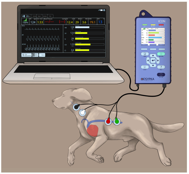
In animals, invasive pulmonary artery thermodilution (PATD) is a gold standard for cardiac output (CO) monitoring, but it is impractical in clinical settings. This study evaluates the agreement between PATD and noninvasive electrical cardiometry (EC) for measuring CO and analyzes the other EC-derived hemodynamic variables in six healthy anesthetized dogs subjected to four different hemodynamic events in a sequential order: (1) euvolemia (baseline); (2) hemorrhage (33% blood volume loss); (3) autologous blood transfusion; and (4) 20 mL/kg colloid bolus. The CO measurements obtained using PATD and EC are compared using Bland-Altman analysis, Lin's concordance correlation (LCC), and polar plot analysis. Values of p < 0.05 are considered significant. The EC measurements consistently underpredict the CO values as compared with PATD, and the LCC is 0.65. The EC's performance is better during hemorrhage, thus indicating its capability in detecting absolute hypovolemia in clinical settings. Even though the percentage error exhibited by EC is 49.4%, which is higher than the standard (<30%), EC displays a good trending ability. Additionally, the EC-derived variables display a significant correlation with the CO measured using PATD. Noninvasive EC may have a potential in monitoring trends in hemodynamics in clinical settings.
Keywords: anesthesia; blood transfusion; canine; colloids; electrical velocimetry; hemodynamics; hemorrhage; hypovolemia; monitoring; noninvasive.
Conflict of interest statement
The authors declare no conflict of interest.
Figures


Similar articles
-
Evaluation of Electrical Cardiometry for Measuring Cardiac Output and Derived Hemodynamic Variables in Comparison with Lithium Dilution in Anesthetized Dogs.Animals (Basel). 2023 Jul 20;13(14):2362. doi: 10.3390/ani13142362. Animals (Basel). 2023. PMID: 37508139 Free PMC article.
-
Performance of four cardiac output monitoring techniques vs. intermittent pulmonary artery thermodilution during a modified passive leg raise maneuver in isoflurane-anesthetized dogs.Front Vet Sci. 2023 Sep 14;10:1238549. doi: 10.3389/fvets.2023.1238549. eCollection 2023. Front Vet Sci. 2023. PMID: 37781276 Free PMC article.
-
Electrical cardiometry for non-invasive cardiac output monitoring: a method comparison study in patients after coronary artery bypass graft surgery.J Clin Monit Comput. 2025 Apr;39(2):371-376. doi: 10.1007/s10877-024-01246-y. Epub 2024 Dec 11. J Clin Monit Comput. 2025. PMID: 39661264 Free PMC article.
-
Accuracy and precision of non-invasive cardiac output monitoring by electrical cardiometry: a systematic review and meta-analysis.J Clin Monit Comput. 2020 Jun;34(3):433-460. doi: 10.1007/s10877-019-00330-y. Epub 2019 Jun 7. J Clin Monit Comput. 2020. PMID: 31175501 Free PMC article.
-
New Developments in Hemodynamic Monitoring.J Cardiothorac Vasc Anesth. 2019 Aug;33 Suppl 1:S67-S72. doi: 10.1053/j.jvca.2019.03.043. J Cardiothorac Vasc Anesth. 2019. PMID: 31279355 Review.
Cited by
-
Evaluation of Electrical Cardiometry for Measuring Cardiac Output and Derived Hemodynamic Variables in Comparison with Lithium Dilution in Anesthetized Dogs.Animals (Basel). 2023 Jul 20;13(14):2362. doi: 10.3390/ani13142362. Animals (Basel). 2023. PMID: 37508139 Free PMC article.
-
Performance of four cardiac output monitoring techniques vs. intermittent pulmonary artery thermodilution during a modified passive leg raise maneuver in isoflurane-anesthetized dogs.Front Vet Sci. 2023 Sep 14;10:1238549. doi: 10.3389/fvets.2023.1238549. eCollection 2023. Front Vet Sci. 2023. PMID: 37781276 Free PMC article.
-
Real-Time Monitoring of Cardiac Output Using Non-Invasive Impedance Cardiography in Dogs: A Pilot Study on Heartworm Extraction and Gastric Decompression.Vet Sci. 2025 May 15;12(5):478. doi: 10.3390/vetsci12050478. Vet Sci. 2025. PMID: 40431571 Free PMC article.
-
Comparison of noninvasive electrical cardiometry and transpulmonary thermodilution for cardiac output measurement in critically ill patients: a prospective observational study.BMC Anesthesiol. 2025 Mar 13;25(1):123. doi: 10.1186/s12871-025-03005-1. BMC Anesthesiol. 2025. PMID: 40082773 Free PMC article.
-
A Comparison of Dobutamine, Norepinephrine, Vasopressin, and Hetastarch for the Treatment of Isoflurane-Induced Hypotension in Healthy, Normovolemic Dogs.Animals (Basel). 2023 Aug 19;13(16):2674. doi: 10.3390/ani13162674. Animals (Basel). 2023. PMID: 37627465 Free PMC article.
References
-
- Mutoh T., Nishimura R., Kim H.Y., Matsunaga S., Sasaki N. Cardiopulmonary effects of sevoflurane, compared with halothane, enflurane, and isoflurane, in dogs. Am. J. Vet. Res. 1997;58:885–890. - PubMed
Grants and funding
LinkOut - more resources
Full Text Sources
Medical
