Review
. 2023 Mar 23;12(4):787.
doi: 10.3390/antiox12040787.

Antioxidant Biomaterials in Cutaneous Wound Healing and Tissue Regeneration: A Critical Review

Affiliations
Review

Antioxidant Biomaterials in Cutaneous Wound Healing and Tissue Regeneration: A Critical Review

Nur Izzah Md Fadilah et al. Antioxidants (Basel). .

Abstract

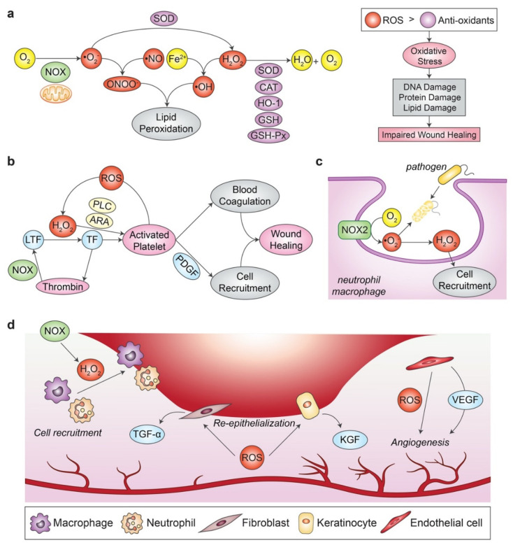
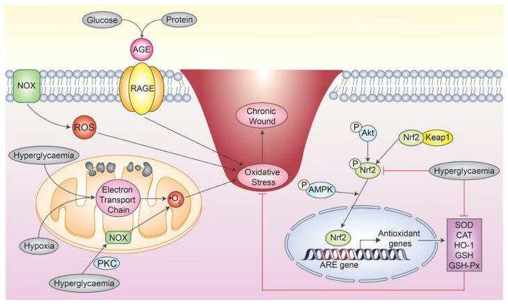
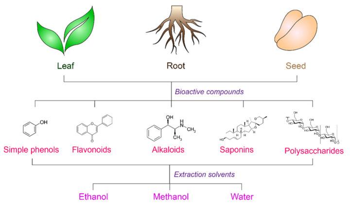
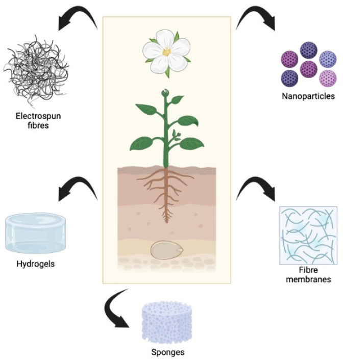
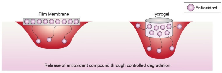
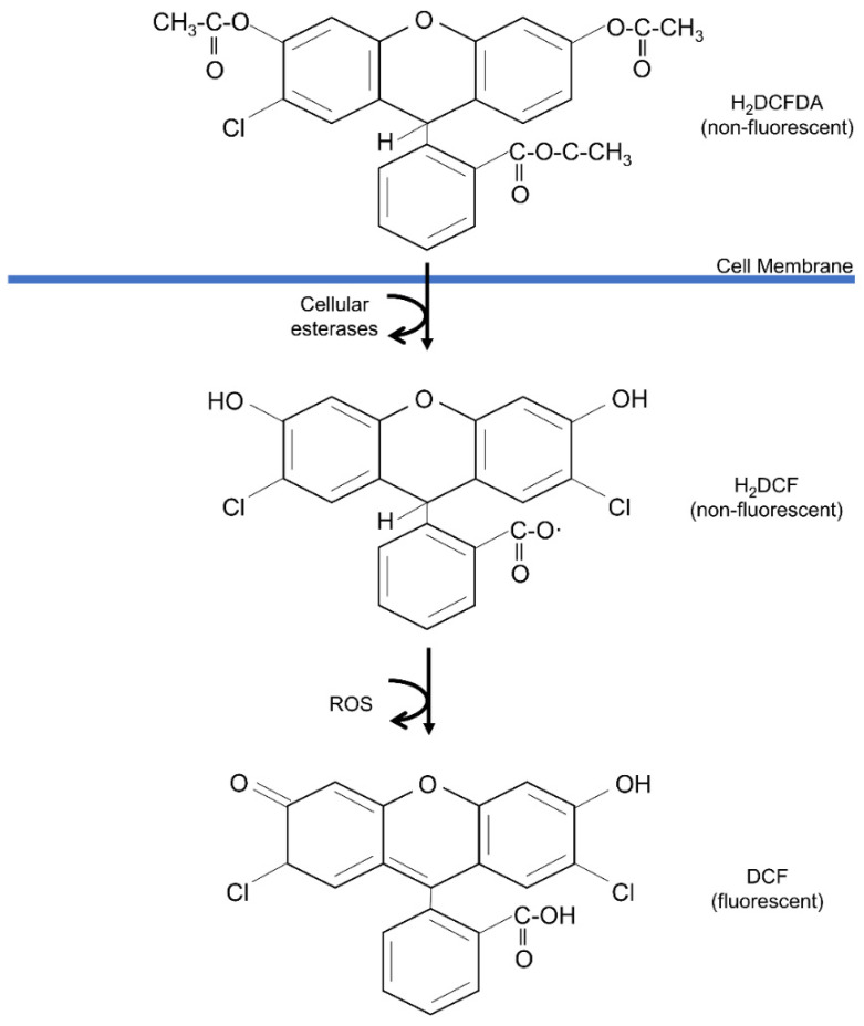
Natural-based biomaterials play an important role in developing new products for medical applications, primarily in cutaneous injuries. A large panel of biomaterials with antioxidant properties has revealed an advancement in supporting and expediting tissue regeneration. However, their low bioavailability in preventing cellular oxidative stress through the delivery system limits their therapeutic activity at the injury site. The integration of antioxidant compounds in the implanted biomaterial should be able to maintain their antioxidant activity while facilitating skin tissue recovery. This review summarises the recent literature that reported the role of natural antioxidant-incorporated biomaterials in promoting skin wound healing and tissue regeneration, which is supported by evidence from in vitro, in vivo, and clinical studies. Antioxidant-based therapies for wound healing have shown promising evidence in numerous animal studies, even though clinical studies remain very limited. We also described the underlying mechanism of reactive oxygen species (ROS) generation and provided a comprehensive review of ROS-scavenging biomaterials found in the literature in the last six years.

Keywords: antioxidant; biomaterials; delivery system; oxidative stress; wound healing.


Conflict of interest statement

The authors declare no conflict of interest.

Figures

Figure 1
Figure 1
ROS and their role in regulating the process of normal wound healing. (a) •O2 can be derived from O2 via the activity of NOX enzyme or the mitochondrial electron transport chain. Next, •O2 can either form ONOO- by reacting with •NO, or they can be converted into H2O2 via SOD activity. H2O2 can further react with Fe2+ to generate •OH, which combines with ONOO and causes lipid peroxidation. Hence, excessive H2O2 are normally eliminated by antioxidants like SOD, CAT, HO-1, GSH, GSH-Px by a reduction reaction into oxygen and water molecule. (b) LTF is activated by H2O2, which leads to thrombin synthesis and platelet activation. The generated thrombin in turn enhances ROS generation via the NOX enzyme, leading to a thrombogenic cycle via ROS-dependent signalling. The activated platelets promote blood coagulation to prevent excessive blood loss and recruit inflammatory cells by secreting PDGF, which altogether results in wound healing. (c) Potential pathogens that invade the wound bed are eliminated via the “respiratory burst” event in phagocytes like neutrophil or macrophage. These cells exhibit an upregulated NOX2 expression upon wound induction, which produces ROS as a weapon to kill the pathogens. (d) H2O2 produced from the NOX enzyme helps in inflammatory cells recruitment to the wound site. ROS production also enhances the re-epithelialisation process by promoting the TGF-α and KGF production in fibroblasts and keratinocytes, respectively. ROS also promote angiogenesis by the activation of endothelial cells via VEGF signalling. ARA: arachidonic acid; CAT: catalase; Fe2+: ferrous ion; GSH: glutathione; GSH-Px: glutathione peroxidase; HO-1: heme oxygenase-1; H2O2: hydrogen peroxide; KGF: keratinocyte growth factor; LTF: latent tissue factor; •NO: nitric oxide radicals; NOX: nicotinamide adenine dinucleotide phosphate (NADPH) oxidase; O2: molecular oxygen; •O2: superoxide anion; •OH: hydroxyl radical; ONOO: peroxynitrite ion; PDGF: platelet-derived growth factor; PLC: phospholipase C; ROS: reactive oxygen species; SOD: superoxide dismutase; TF: tissue factor; TGF-α: transforming growth factor-alpha; VEGF: vascular endothelial growth factor. The idea of the figure is adapted from [49,50,51].
Figure 2
Figure 2
Oxidative stress underlying a chronic wound. Excessive ROS are produced via the mitochondrial electron transport chain under hyperglycaemia and hypoxic condition. Hyperglycaemia can also activate mitochondrial NOX activity via PKC. Next, the formation of AGE from the reaction between glucose and protein binds to its receptor, RAGE, which induces signalling that leads to oxidative stress. Besides promoting the formation of oxidants, hyperglycaemia also inhibits the Nrf2 signalling, which leads to downregulated expression of antioxidants. Altogether, the persistent redox imbalance leads to severe oxidative stress and results in a chronic wound. AMPK: adenosine monophosphate-activated protein kinase; AGE: advanced glycation end product; ARE: antioxidant response element; CAT: catalase; GSH: glutathione; GSH-Px: glutathione peroxidase; HO-1: heme oxygenase-1; Keap1: Kelch-like ECH-associated protein 1; NOX: nicotinamide adenine dinucleotide phosphate (NADPH) oxidase; •O2: superoxide anion; Nrf2: nuclear factor erythroid 2-related factor 2; PKC: protein kinase C; RAGE: AGE receptor; ROS: reactive oxygen species; SOD: superoxide dismutase. The idea of the figure is adapted from [54,55,56].
Figure 3
Figure 3
The selected plant parts, bioactive structures, and primary extractions of antioxidants are involved in wound healing. The idea of the figure is adapted from [57].
Figure 4
Figure 4
Different types of materials from plant-derived antioxidant compounds. The idea of the figure is adapted from [104].
Figure 5
Figure 5
Control release antioxidant compounds from biomaterials. The idea of the figure is adapted from [169].
Figure 6
Figure 6
The reaction of DPPH assay when the antioxidant agent is present. The image is adapted from Marjoni and Zulfisa (2017) [172], licensed under Creative Commons Attribution License.
Figure 7
Figure 7
The interaction of ABTS assay when the antioxidant agent is present. The image is adapted from Bedlovicova et al. [174] and license under CC BY 4.0.
Figure 8
Figure 8
The chain reaction of DCFDA assay where the molecules become highly fluorescent after reacting with reactive oxygen species. The image is adapted from Nova et al. [176] and license under CC BY 4.0.

References

    1. Zhang F., Han X., Guo C., Yang H., Wang J., Wu X. Fibrous aramid hydrogel supported antibacterial agents for accelerating bacterial-infected wound healing. Mater. Sci. Eng. C. 2021;121:1–10. doi: 10.1016/j.msec.2020.111833. - DOI - PubMed
    1. Naseri-Nosar M., Ziora Z.M. Wound dressings from naturally-occurring polymers: A review on homopolysaccharide-based composites. Carbohydr. Polym. 2018;189:379–398. doi: 10.1016/j.carbpol.2018.02.003. - DOI - PubMed
    1. Yousef H., Alhajj M., Sharma S. Anatomy, Skin (Integument), Epidermis-StatPearls-NCBI Bookshelf. StatPearls Publishing; Treasure Island, FL, USA: 2020. pp. 1–12.
    1. Nike D.U., Md Fadilah N.I., Sallehuddin N., Azlan A.Y.H.N., Imran F.H., Maarof M., Fauzi M.B. Genipin-Crosslinking Effects on Biomatrix Development for Cutaneous Wound Healing: A Concise Review. Front. Bioeng. Biotechnol. 2022;10:726. - PMC - PubMed
    1. Boateng J., Catanzano O. Advanced therapeutic dressings for effective wound healing—A review. J. Pharm. Sci. 2015;104:3653–3680. doi: 10.1002/jps.24610. - DOI - PubMed

LinkOut - more resources