. 2023 Apr 8;12(4):900.
doi: 10.3390/antiox12040900.

Mitochondrial ROS Triggers KIN Pathogenesis in FAN1-Deficient Kidneys

Affiliations

Mitochondrial ROS Triggers KIN Pathogenesis in FAN1-Deficient Kidneys

Merlin Airik et al. Antioxidants (Basel). .

Abstract

Karyomegalic interstitial nephritis (KIN) is a genetic adult-onset chronic kidney disease (CKD) characterized by genomic instability and mitotic abnormalities in the tubular epithelial cells. KIN is caused by recessive mutations in the FAN1 DNA repair enzyme. However, the endogenous source of DNA damage in FAN1/KIN kidneys has not been identified. Here we show, using FAN1-deficient human renal tubular epithelial cells (hRTECs) and FAN1-null mice as a model of KIN, that FAN1 kidney pathophysiology is triggered by hypersensitivity to endogenous reactive oxygen species (ROS), which cause chronic oxidative and double-strand DNA damage in the kidney tubular epithelial cells, accompanied by an intrinsic failure to repair DNA damage. Furthermore, persistent oxidative stress in FAN1-deficient RTECs and FAN1 kidneys caused mitochondrial deficiencies in oxidative phosphorylation and fatty acid oxidation. The administration of subclinical, low-dose cisplatin increased oxidative stress and aggravated mitochondrial dysfunction in FAN1-deficient kidneys, thereby exacerbating KIN pathophysiology. In contrast, treatment of FAN1 mice with a mitochondria-targeted ROS scavenger, JP4-039, attenuated oxidative stress and accumulation of DNA damage, mitigated tubular injury, and preserved kidney function in cisplatin-treated FAN1-null mice, demonstrating that endogenous oxygen stress is an important source of DNA damage in FAN1-deficient kidneys and a driver of KIN pathogenesis. Our findings indicate that therapeutic modulation of kidney oxidative stress may be a promising avenue to mitigate FAN1/KIN kidney pathophysiology and disease progression in patients.

Keywords: DNA damage; FAN1; chronic kidney disease; karyomegalic interstitial nephritis; oxidative stress.


Conflict of interest statement

The authors declare no conflict of interest. The funders had no role in the design of the study; in the collection, analyses, or interpretation of data; in the writing of the manuscript; or in the decision to publish the results.

Figures

Figure 1
Figure 1
KIN is associated with impaired oxidative phosphorylation and fatty acid oxidation. (A) Gene ontology analysis of control and FAN1KO kidneys after repeated low-dose cisplatin injury. The graph shows −log p values calculated using the Benjamini–Hochberg-corrected two-tailed t-test for the enrichment of a specific pathway. (B) Gene-set enrichment signatures of oxidative phosphorylation genes in cisplatin-treated FAN1KO vs. control kidneys. Normalized Enrichment Score (NES) −2.285536, p < 0.0001, FDR q-value 0.0001. (C) Gene-set enrichment signatures of fatty acid metabolism genes in cisplatin-treated FAN1KO vs. control kidneys. Normalized Enrichment Score (NES) −2.754839, p < 0.0001, FDR q-value 0.0001. (D) qPCR analysis of Ndufa1, Sdha1, and Cox4l1 mRNA expression. (ctrl 1.6 ± 0.3 vs. FAN1KO 6.9 ± 0.6, * p < 0.05, ** p < 0.01; *** p < 0.001), n = 5 each. (E) Western blot analysis of ATP5a, UQRC2, MTCO1, SDHB, and NDUFB8 using rodent OXPHOS mitococtail. Ponceau staining shows equal protein loading. (F) qPCR analysis of Acadm, Acadvl, Cpt2, and Echs1 mRNA expression. (ctrl 1.6 ± 0.3 vs. FAN1KO 6.9 ± 0.6, * p < 0.05, ** p < 0.01; *** p < 0.001), n = 5 each. (DF) Data are presented as the mean ± SEM. A two-way ANOVA with Tukey’s post hoc analysis.
Figure 2
Figure 2
KIN pathogenesis is associated with increased lipid peroxidation, accumulation of neutral lipids, and oxidative stress in the kidney proximal tubule cells. (A) Representative images of cisplatin-treated control and FAN1KO kidneys stained with antibodies against 4-HNE to mark lipid peroxidation products and Lotus tetragonolobus lectin (LTL), which marks proximal tubule cells. **** p < 0.0001, n = 4 each. Scale bars: 40 μm. (B) Representative images of cisplatin-treated control and FAN1KO kidneys stained with OilRed O. *** p < 0.001, n = 4 each. Scale bars 50 μm. (C) Quantification of 8-OHdG staining in LTL-positive proximal tubules in untreated and cisplatin-treated wild-type and FAN1KO kidneys. ** p < 0.01, MFI—mean fluorescent intensity. (D) Quantification of CM-H2DCFDA staining in untreated and cisplatin-treated wild-type and FAN1KO kidneys. * p < 0.05, MFI—mean fluorescent intensity. Scale bars: 50 mm. (AD) Data are presented as the mean ± SEM. A two-way ANOVA with Tukey’s post hoc analysis.
Figure 3
Figure 3
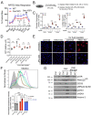
FAN1KO renal tubular epithelial cells have impaired mitochondrial metabolism and increased ROS production. (A) Representative oxygen consumption traces for parental (WT) and FAN1KO hRTEC mitochondria using Oroboros high-resolution respirometry. Basal, baseline respiration; CI, Electron Transport Complex I; CII, Electron Transport Complex II; Mal, malate; ADP, adenosine diphosphate; Pyr, pyruvate; Glut, glutamate; Succ, succinate; CCCP, carbonyl cyanide m-chlorophenylhydrazone; Rot, rotenone. Rotenone (CI) inhibitor was used to separate the contributions of complexes I and II. (B) Overview of cisplatin and JP4-039 treatment protocols in FAN1KO human renal tubular epithelial cells. Cells were seeded for 24 h and left untreated or treated with 5 μM cisplatin for 1 h, followed by two rises with PBS and replaced with fresh media. When JP4-039 treatment was performed following cisplatin treatment, cells were daily provided with fresh media ±JP4-039 (5 μM) for the duration of the culture period. (C) Glucose consumption and lactose secretion measurements. Parental and FAN1KO hRTECs were ± treated with cisplatin, the media were harvested, and glucose consumption and lactate production were quantified (n = 6). (D) ATP measurement in parental and FAN1KO hRTECs at indicated time points after treatment with cisplatin. Data are shown after normalizing ATP levels in untreated parental cells. N = 3 independent experiments. (E) Representative images of MitoSox staining in parental and FAN1KO hRTECs. Cells were ± treated with 5 μM cisplatin for 1 h, followed by ±JP4-039 (5 μM) for 48 h. (F) Detection of mitochondrial ROS (MitoSox) by flow cytometry in parental and FAN1KO hRTECs. Cells were ± treated with 5 μM cisplatin for 1 h, followed by ±JP4-039 (5 μM) for 72 h. * p < 0.05, ** p < 0.01, **** p < 0.0001, n = 3 independent experiments. (G) Representative Western blot analysis of pATR, FANCD2, CDC6, pRPA32, pCHK1, and γH2AX in FAN1KO hRTECs. Western blot analysis of DNA repair pathway markers in hRTEC chromatin preparation reveals the activation of the Fanconi anemia repair pathway (ubiquitination of FANCD2, marked by a star), increased levels of replication stress (pRPA32 S4/S8), and DNA double-strand breaks (γH2AX) in cisplatin-treated FAN1KO cells. Histone H3 is used as a chromatin loading control. Cells were ± treated with 5 μM cisplatin for 1 h followed by ±JP4-039 (5 μM) for 48 h. (A,C,E) Data are presented as the mean ± SEM. (A) A two-way ANOVA with Tukey’s post hoc analysis. (C,D) Ordinary one-way ANOVA with Tukey’s post hoc analysis.
Figure 4
Figure 4
DNA damage causes aberrant cell cycle activity in FAN1-null kidneys. (A) Mice received 2 doses of intraperitoneal (i.p.) cisplatin (2 mg/kg) vs. vehicle (normal saline) on days 0 and 7 and were treated with i.p. JP4-039 (10 mg/kg in 50% PEG-400/50% H2O) vs. vehicle (50% PEG-400/50% H2O) 1 day prior to and until day 14 after the first cisplatin injection. Tissues were collected for analysis 21 days after the first cisplatin dose. (B) Blood urea nitrogen (BUN) measurements in ctrl and FAN1-null mice show loss of kidney function in FAN1KO mice after induction of KIN. *** p < 0.001, **** p < 0.0001, n = 5 each. (C) Histological analysis of kidney sections by Periodic acid–Schiff (PAS) staining demonstrates the formation of KIN in FAN1KO mice, characterized by tubular atrophy, formation of karyomegalic nuclei (red arrowheads), and segmental basement membrane thickening (black arrowheads) in the proximal tubules. Scale bars: 50 μm. (D) Tubular injury scores in control mice were compared with those in FAN1KO kidneys after low-dose cisplatin administration. ** p < 0.01, *** p < 0.001, **** p < 0.0001, n = 5 each. (B,D) Data are presented as the mean ± SEM. A two-way ANOVA with Tukey’s post hoc analysis.
Figure 5
Figure 5
Oxidation stress-induced tubular damage is attenuated by JP4-039. (A) Representative images of untreated and cisplatin (2 × 2 mg/kg) ±JP4-039 (10 mg/kg in 50% PEG-400/50% H2O) treated control and FAN1KO kidneys stained with antibodies against 4-HNE to mark lipid peroxidation products. *** p < 0.001, n = 4 each. Scale bar: 25 μm. (B) Increased lipotoxicity in injured FAN1KO kidneys as revealed by OilRed O staining. Treatment with JP4-039 reduces lipid accumulation in cisplatin-injured FAN1KO kidneys. **** p < 0.0001, n = 5 each. Scale bar: 50 μm. (A,B) Data are presented as the mean ± SEM. A two-way ANOVA with Tukey’s post hoc analysis.

References

    1. Zhou W., Otto E.A., Cluckey A., Airik R., Hurd T.W., Chaki M., Diaz K., Lach F.P., Bennett G.R., Gee H.Y., et al. FAN1 mutations cause karyomegalic interstitial nephritis, linking chronic kidney failure to defective DNA damage repair. Nat. Genet. 2012;44:910–915. doi: 10.1038/ng.2347. - DOI - PMC - PubMed
    1. Airik R., Schueler M., Airik M., Cho J., Porath J.D., Mukherjee E., Sims-Lucas S., Hildebrandt F. A FANCD2/FANCI-Associated Nuclease 1-Knockout Model Develops Karyomegalic Interstitial Nephritis. J. Am. Soc. Nephrol. 2016;27:3552–3559. doi: 10.1681/ASN.2015101108. - DOI - PMC - PubMed
    1. Spoendlin M., Moch H., Brunner F., Brunner W., Burger H.R., Kiss D., Wegmann W., Dalquen P., Oberholzer M., Thiel G., et al. Karyomegalic interstitial nephritis: Further support for a distinct entity and evidence for a genetic defect. Am. J. Kidney Dis. 1995;25:242–252. doi: 10.1016/0272-6386(95)90005-5. - DOI - PubMed
    1. Dash J., Saudan P., Paoloni-Giacobino A., Moll S., de Seigneux S. Case report: A 58-year-old man with small kidneys and elevated liver enzymes. BMC Nephrol. 2020;21:107. doi: 10.1186/s12882-020-01762-4. - DOI - PMC - PubMed
    1. Gueguen L., Delaval R., Blanluet M., Sartelet H., Leou S., Dubois d’Enghien C., Golmard L., Stoppa-Lyonnet D., Testevuide P., Faguer S. Recurrent FAN1 p.W707X Pathogenic Variant Originating Before ad 1800 Underlies High Frequency of Karyomegalic Interstitial Nephritis in South Pacific Islands. Kidney Int. Rep. 2021;6:2207–2211. doi: 10.1016/j.ekir.2021.05.010. - DOI - PMC - PubMed