. 2023 Mar 31;14(4):845.
doi: 10.3390/genes14040845.

Damage-Free Shortening of Telomeres Is a Potential Strategy Supporting Blind Mole-Rat Longevity

Affiliations

Damage-Free Shortening of Telomeres Is a Potential Strategy Supporting Blind Mole-Rat Longevity

Huda Adwan Shekhidem et al. Genes (Basel). .

Abstract

Telomere shortening or loss of shelterin components activates DNA damage response (DDR) pathways, leading to a replicative senescence that is usually coupled with a senescence-associated secretory phenotype (SASP). Recent studies suggested that telomere aberration that activates DDR may occur, irrespective of telomere length or loss of shelterin complex. The blind mole-rat (Spalax) is a subterranean rodent with exceptional longevity, and its cells demonstrate an uncoupling of senescence and SASP inflammatory components. Herein, we evaluated Spalax relative telomere length, telomerase activity, and shelterin expression, along with telomere-associated DNA damage foci (TAFs) levels with cell passage. We show that telomeres shorten in Spalax fibroblasts similar to the process in rats, and that the telomerase activity is lower. Moreover, we found lower DNA damage foci at the telomeres and a decline in the mRNA expression of two shelterin proteins, known as ATM/ATR repressors. Although additional studies are required for understanding the underling mechanism, our present results imply that Spalax genome protection strategies include effective telomere maintenance, preventing early cellular senescence induced by persistent DDR, thereby contributing to its longevity and healthy aging.

Keywords: DNA damage; Spalax; senescence; shelterin; telomerase activity; telomere length.


Conflict of interest statement

The authors declare no conflict of interest.

Figures

Figure 1
Figure 1
Relative telomere length (telomere to single copy gene (T/S) ratio) and relative telomerase activity in Spalax and rats’ primary fibroblasts. (a) Relative telomere length (rTL) as a function of passages in Spalax primary fibroblasts (slope = −0.7750, F1,7 = 12.6, p < 0.01, R2 = 0.6429); (b) relative telomere length (rTL) as a function of passages in rat primary fibroblast cells (slope = −0.2793, F1,7 = 6.969, p < 0.05, R2 = 0.4989) (Pearson’s); (c) range of rTL between Spalax and rat fibroblasts (boxplots represent data from a,b); (d) range of relative telomerase activity (rTA) between Spalax and rat fibroblasts (both passages); (e) rTA in Spalax and rat primary fibroblast cells with cell passage (n = 3, cells from three different individuals of Spalax and rats). ** p < 0.01 and *** p < 0.001 (Mann–Whitney U test).
Figure 2
Figure 2
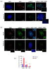
TAFs formation in Spalax and rat cells. Immunostaining for TRF2 (red) and γ-H2A.X (green) cell nuclei were counterstained with DAPI (blue) for early-passage cells (a) and late-passage, senescent cells (b). Examples of double stained foci are marked with rectangles. (c) The average number of γ-H2A.X, TRF2 and TAFs (co-localized) foci counted in 20 randomly chosen nuclei with γ-H2A.X positive foci using FociCounter. *** p < 0.001 (Mann–Whitney U-test).
Figure 3
Figure 3
Levels of relative transcription of shelterin complex core components (af) in primary fibroblast cells of Spalax and rats. The mRNA expression rates were quantified in three different passages; passage 2, 4, and 5 (P2, P4 and P5) by using qRT–PCR. Relative transcription levels are presented as mean ± SD of three independent experiments (n = 3, cells from three different individuals of Spalax and rats). * p < 0.05, ** p < 0.01, and *** p < 0.001 (Mann–Whitney U test). Note that the relative standard curve was built for each species separately; therefore, data points of each species can only be compared to its control (1.0). The differences between P4 and P5 were insignificant.

References

    1. Zhu Y., Liu X., Ding X., Wang F., Geng X. Telomere and its role in the aging pathways: Telomere shortening, cell senescence and mitochondria dysfunction. Biogerontology. 2018;20:1–16. doi: 10.1007/s10522-018-9769-1. - DOI - PubMed
    1. Birch J., Barnes P.J., Passos J.F. Mitochondria, telomeres and cell senescence: Implications for lung ageing and disease. Pharmacol. Ther. 2017;183:34–49. doi: 10.1016/j.pharmthera.2017.10.005. - DOI - PubMed
    1. Bernadotte A., Mikhelson V.M., Spivak I.M. Markers of cellular senescence. Telomere shortening as a marker of cellular senescence. Aging. 2016;8:3–11. doi: 10.18632/aging.100871. - DOI - PMC - PubMed
    1. van Deursen J.M. The role of senescent cells in ageing. Nature. 2014;509:439–446. doi: 10.1038/nature13193. - DOI - PMC - PubMed
    1. O’Sullivan R.J., Karlseder J. Telomeres: Protecting chromosomes against genome instability. Nat. Rev. Mol. Cell Biol. 2010;11:171–181. doi: 10.1038/nrm2848. - DOI - PMC - PubMed

Publication types