

Surveillance of cfDNA Hot Spot Mutations in NSCLC Patients during Disease Progression
- PMID: 37108122
- PMCID: PMC10138687
- DOI: 10.3390/ijms24086958
Surveillance of cfDNA Hot Spot Mutations in NSCLC Patients during Disease Progression
Abstract
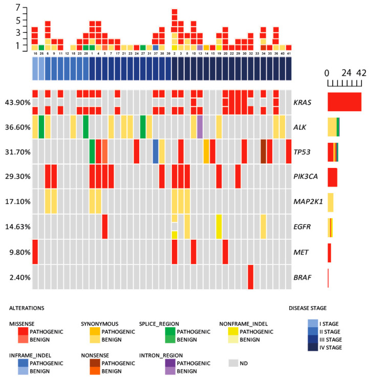
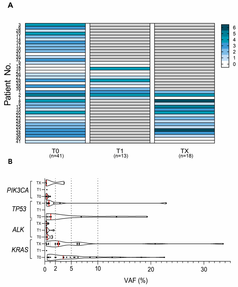
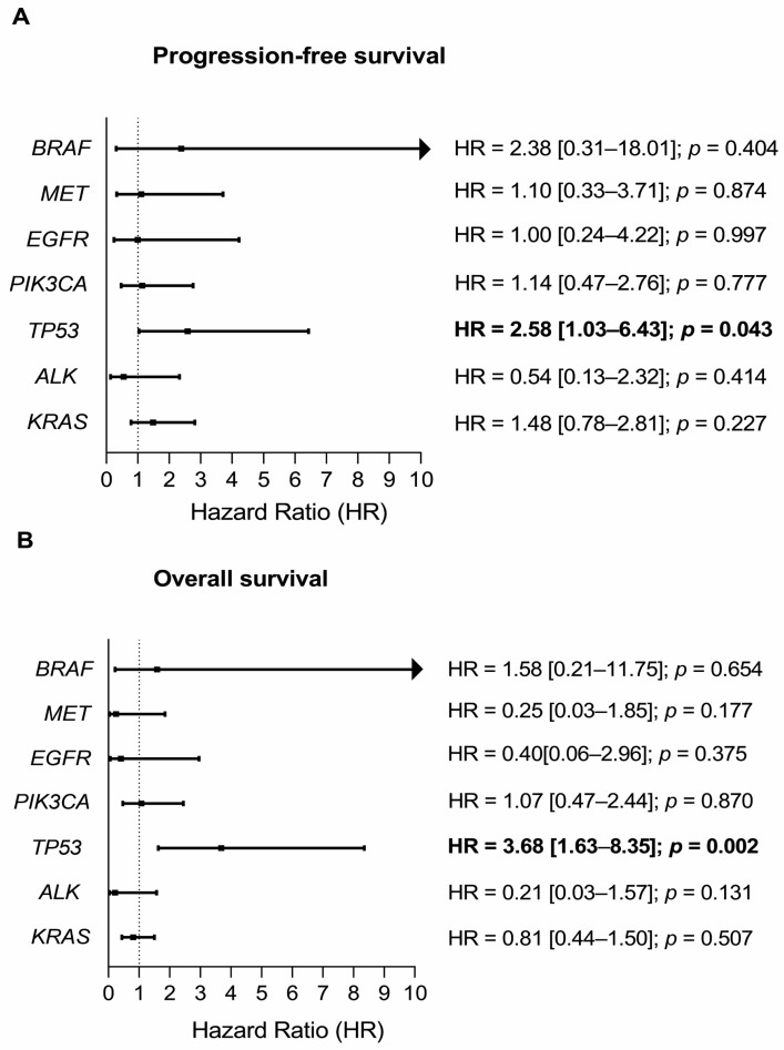
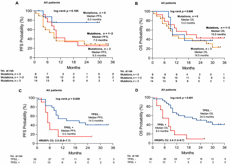
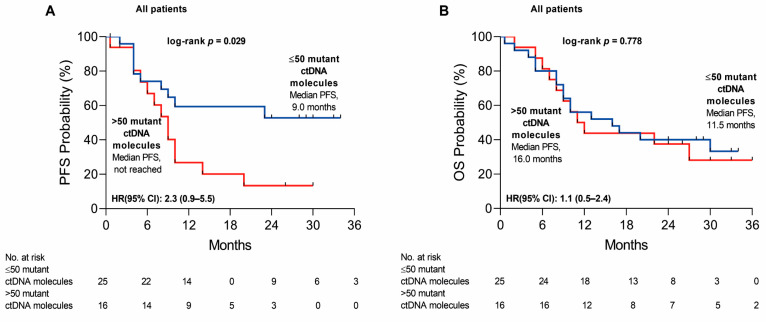
Non-small cell cancer (NSCLC) has been identified with a great variation of mutations that can be surveyed during disease progression. The aim of the study was to identify and monitor lung cancer-specific mutations incidence in cell-free DNA as well as overall plasma cell-free DNA load by means of targeted next-generation sequencing. Sequencing libraries were prepared from cell-free DNA (cfDNA) isolated from 72 plasma samples of 41 patients using the Oncomine Lung cfDNA panel covering hot spot regions of 11 genes. Sequencing was performed with the Ion Torrent™ Ion S5™ system. Four genes were detected with highest mutation incidence: KRAS (43.9% of all cases), followed by ALK (36.6%), TP53 (31.7%), and PIK3CA (29.3%). Seven patients had co-occurring KRAS + TP53 (6/41, 14.6%) or KRAS + PIK3CA (7/41, 17.1%) mutations. Moreover, the mutational status of TP53 as well an overall cell-free DNA load were confirmed to be predictors of poor progression-free survival (HR = 2.5 [0.8-7.7]; p = 0.029 and HR = 2.3 [0.9-5.5]; p = 0.029, respectively) in NSCLC patients. In addition, TP53 mutation status significantly predicts shorter overall survival (HR = 3.4 [1.2-9.7]; p < 0.001). We demonstrated that TP53 mutation incidence as well as a cell-free DNA load can be used as biomarkers for NSCLC monitoring and can help to detect the disease progression prior to radiological confirmation of the status.
Keywords: KRAS; TP53; cell-free DNA (cfDNA) mutation load; liquid biopsy; non-small cell lung cancer (NSCLC).
Conflict of interest statement
The authors declare no conflict of interest.
Figures
Similar articles
-
Cross-platform comparison of next-generation sequencing and matrix-assisted laser desorption/ionization time-of-flight mass spectrometry for detecting KRAS/NRAS/BRAF/PIK3CA mutations in cfDNA from metastatic colorectal cancer patients.J Clin Lab Anal. 2021 Sep;35(9):e23818. doi: 10.1002/jcla.23818. Epub 2021 Aug 17. J Clin Lab Anal. 2021. PMID: 34403504 Free PMC article.
-
Role of circulating free DNA in evaluating clinical tumor burden and predicting survival in Chinese metastatic colorectal cancer patients.BMC Cancer. 2020 Oct 16;20(1):1006. doi: 10.1186/s12885-020-07516-7. BMC Cancer. 2020. PMID: 33066758 Free PMC article.
-
The potential of PIK3CA, KRAS, BRAF, and APC hotspot mutations as a non-invasive detection method for colorectal cancer.Mol Cell Probes. 2022 Jun;63:101807. doi: 10.1016/j.mcp.2022.101807. Epub 2022 Mar 13. Mol Cell Probes. 2022. PMID: 35296442
-
Plasma Cell-Free DNA Genotyping: From an Emerging Concept to a Standard-of-Care Tool in Metastatic Non-Small Cell Lung Cancer.Oncologist. 2021 Oct;26(10):e1812-e1821. doi: 10.1002/onco.13889. Epub 2021 Jul 26. Oncologist. 2021. PMID: 34216176 Free PMC article. Review.
-
KRAS mutation as a prognostic factor and predictive factor in advanced/metastatic non-small cell lung cancer: A systematic literature review and meta-analysis.Cancer Treat Res Commun. 2020;24:100200. doi: 10.1016/j.ctarc.2020.100200. Epub 2020 Jul 25. Cancer Treat Res Commun. 2020. PMID: 32750661
Cited by
-
Next generation sequencing and genomic mapping: towards precision molecular diagnosis of lung cancer in Morocco.Pan Afr Med J. 2024 Nov 13;49:75. doi: 10.11604/pamj.2024.49.75.45306. eCollection 2024. Pan Afr Med J. 2024. PMID: 39989937 Free PMC article.
-
Detecting actionable mutations from matched plasma-based versus tissue next-generation sequencing in advanced non-small cell lung cancer: a retrospective single centre analysis on site.J Exp Clin Cancer Res. 2025 Aug 6;44(1):229. doi: 10.1186/s13046-025-03480-x. J Exp Clin Cancer Res. 2025. PMID: 40765058 Free PMC article.
-
Co-Occurring Genomic Alterations in NSCLC: Making Order into a Crowded List.Cancers (Basel). 2025 Jul 18;17(14):2388. doi: 10.3390/cancers17142388. Cancers (Basel). 2025. PMID: 40723270 Free PMC article. Review.
-
TP53-centric ctDNA complements PET/CT for non-invasive assessment of pathological complete response and survival after neoadjuvant immunochemotherapy in esophageal squamous cell carcinoma: a prospective cohort study.Int J Surg. 2025 May 1;111(5):3256-3268. doi: 10.1097/JS9.0000000000002341. Int J Surg. 2025. PMID: 40146232 Free PMC article.
References
-
- Cancer Today. [(accessed on 7 November 2022)]. Available online: http://gco.iarc.fr/today/home.
MeSH terms
Substances
Grants and funding
LinkOut - more resources
Full Text Sources
Medical
Research Materials
Miscellaneous
