

Improved Wound Healing and Skin Regeneration Ability of 3,2'-Dihydroxyflavone-Treated Mesenchymal Stem Cell-Derived Extracellular Vesicles
- PMID: 37108128
- PMCID: PMC10138514
- DOI: 10.3390/ijms24086964
Improved Wound Healing and Skin Regeneration Ability of 3,2'-Dihydroxyflavone-Treated Mesenchymal Stem Cell-Derived Extracellular Vesicles
Abstract
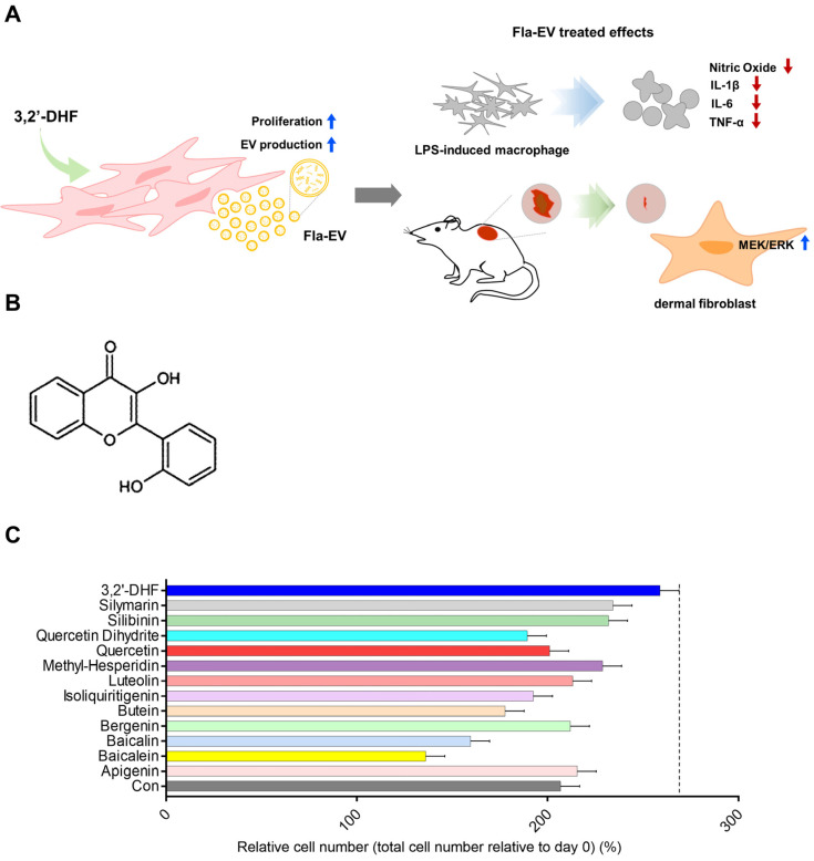
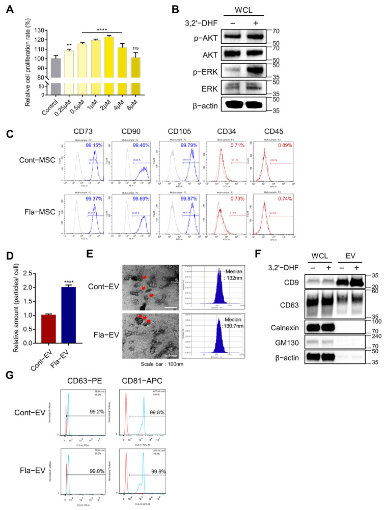
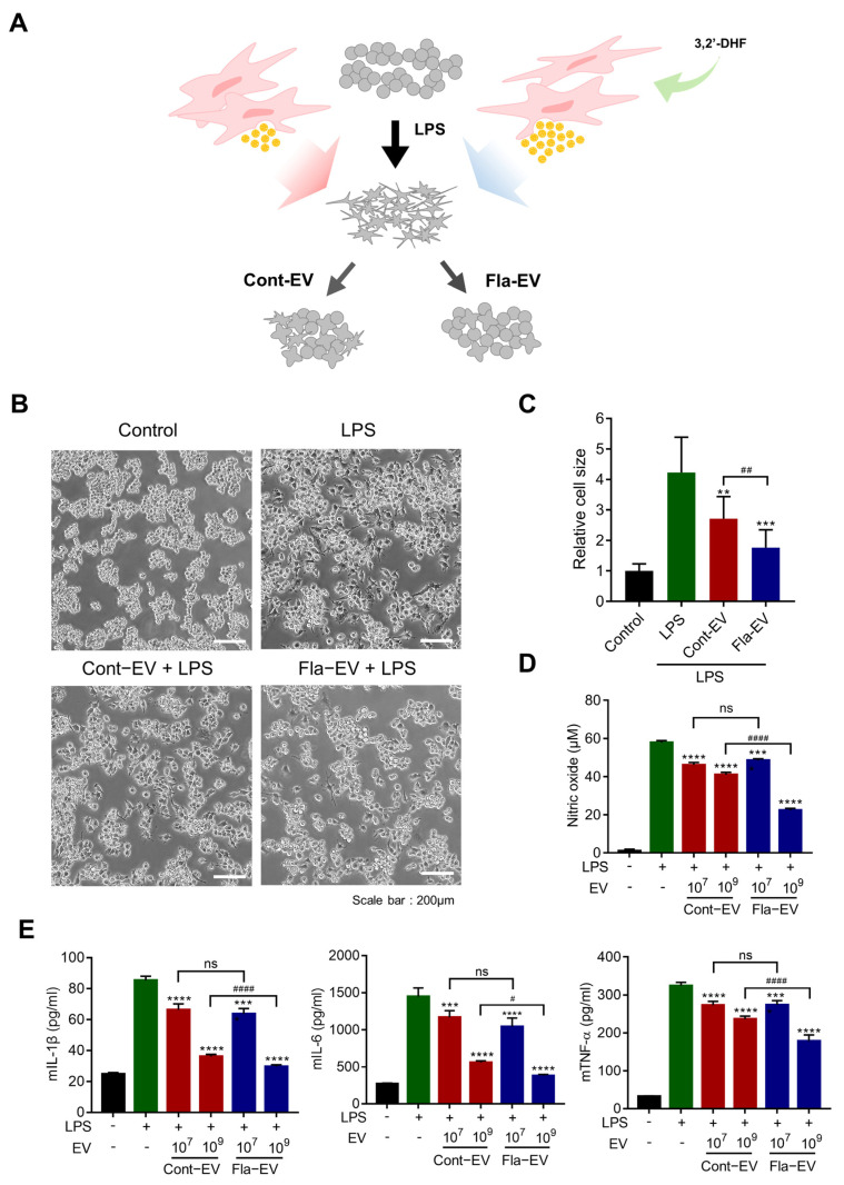
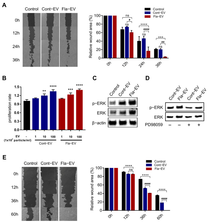
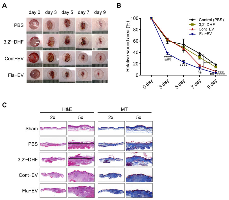
Flavonoids enhance the self-renewal and differentiation potential of mesenchymal stem cells (MSCs) and have therapeutic activities, including regenerative, anti-oxidative, and anti-inflammatory effects. Recent studies have revealed that MSC-derived extracellular vesicles (MSC-EVs) have therapeutic effects on tissue regeneration and inflammation. To facilitate further research on the therapeutic potential of MSC-EVs derived from flavonoid-treated MSCs, we surveyed the production of EVs and their therapeutic applications in wound regeneration. MSCs treated with flavonoids enhanced EV production twofold compared with naïve MSCs. EVs produced by MSCs treated with flavonoids (Fla-EVs) displayed significant anti-inflammatory and wound-healing effects in vitro. The wound-healing capacity of EVs was mediated by the upregulation of mitogen-activated protein kinase kinase (MEK)/extracellular signal-regulated kinase (ERK) signaling. Interestingly, the protein level of p-ERK under inhibition of MEK signals was maintained in Fla-EV-treated fibroblasts, suggesting that Fla-EVs have a higher therapeutic potential than naïve MSC-EVs (Cont-EVs) in wound healing. Moreover, the in vivo wound closure effect of the Fla-EVs showed significant improvement compared with that of the flavonoid-only treatment group and the Cont-EVs. This study provides a strategy for the efficient production of EVs with superior therapeutic potential using flavonoids.
Keywords: EVs; MEK/ERK signal; MSCs; anti-inflammation; flavonoid; wound healing.
Conflict of interest statement
The authors declare no conflict of interest.
Figures
Similar articles
-
Extracellular Vesicles Derived from Mesenchymal Stem Cells Promote Wound Healing and Skin Regeneration by Modulating Multiple Cellular Changes: A Brief Review.Genes (Basel). 2023 Jul 25;14(8):1516. doi: 10.3390/genes14081516. Genes (Basel). 2023. PMID: 37628568 Free PMC article. Review.
-
Roles of extracellular vesicles from mesenchymal stem cells in regeneration.Mol Cells. 2024 Dec;47(12):100151. doi: 10.1016/j.mocell.2024.100151. Epub 2024 Nov 13. Mol Cells. 2024. PMID: 39547584 Free PMC article. Review.
-
Extracellular vesicle-carried microRNA-27b derived from mesenchymal stem cells accelerates cutaneous wound healing via E3 ubiquitin ligase ITCH.J Cell Mol Med. 2020 Oct;24(19):11254-11271. doi: 10.1111/jcmm.15692. Epub 2020 Aug 26. J Cell Mol Med. 2020. PMID: 32845084 Free PMC article.
-
MSC-EVs and UCB-EVs promote skin wound healing and spatial transcriptome analysis.Sci Rep. 2025 Feb 1;15(1):4006. doi: 10.1038/s41598-025-87592-6. Sci Rep. 2025. PMID: 39893214 Free PMC article.
-
Perspectives for Future Use of Extracellular Vesicles from Umbilical Cord- and Adipose Tissue-Derived Mesenchymal Stem/Stromal Cells in Regenerative Therapies-Synthetic Review.Int J Mol Sci. 2020 Jan 25;21(3):799. doi: 10.3390/ijms21030799. Int J Mol Sci. 2020. PMID: 31991836 Free PMC article. Review.
Cited by
-
Efficacy of Allogeneic and Xenogeneic Exosomes for the Treatment of Canine Atopic Dermatitis: A Pilot Study.Animals (Basel). 2024 Jan 16;14(2):282. doi: 10.3390/ani14020282. Animals (Basel). 2024. PMID: 38254451 Free PMC article.
-
Antifungal and antibiofilm activities of flavonoids against Candida albicans: Focus on 3,2'-dihydroxyflavone as a potential therapeutic agent.Biofilm. 2024 Jul 26;8:100218. doi: 10.1016/j.bioflm.2024.100218. eCollection 2024 Dec. Biofilm. 2024. PMID: 39175909 Free PMC article.
-
Antibiofilm and Antivirulence Potentials of 3,2'-Dihydroxyflavone against Staphylococcus aureus.Int J Mol Sci. 2024 Jul 24;25(15):8059. doi: 10.3390/ijms25158059. Int J Mol Sci. 2024. PMID: 39125628 Free PMC article.
-
Mesenchymal stem cells-derived small extracellular vesicles and apoptotic extracellular vesicles for wound healing and skin regeneration: a systematic review and meta-analysis of preclinical studies.J Transl Med. 2025 Mar 24;23(1):364. doi: 10.1186/s12967-024-05744-0. J Transl Med. 2025. PMID: 40128791 Free PMC article.
-
Unveiling the wonders of bacteria-derived extracellular vesicles: From fundamental functions to beneficial applications.Heliyon. 2025 Feb 6;11(4):e42509. doi: 10.1016/j.heliyon.2025.e42509. eCollection 2025 Feb 28. Heliyon. 2025. PMID: 40028522 Free PMC article. Review.
References
MeSH terms
Substances
Grants and funding
LinkOut - more resources
Full Text Sources
Miscellaneous
