

The Potential Use of Mitochondrial Extracellular Vesicles as Biomarkers or Therapeutical Tools
- PMID: 37108168
- PMCID: PMC10139054
- DOI: 10.3390/ijms24087005
The Potential Use of Mitochondrial Extracellular Vesicles as Biomarkers or Therapeutical Tools
Abstract
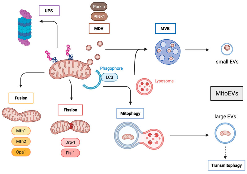
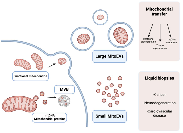
The mitochondria play a crucial role in cellular metabolism, reactive oxygen species (ROS) production, and apoptosis. Aberrant mitochondria can cause severe damage to the cells, which have established a tight quality control for the mitochondria. This process avoids the accumulation of damaged mitochondria and can lead to the release of mitochondrial constituents to the extracellular milieu through mitochondrial extracellular vesicles (MitoEVs). These MitoEVs carry mtDNA, rRNA, tRNA, and protein complexes of the respiratory chain, and the largest MitoEVs can even transport whole mitochondria. Macrophages ultimately engulf these MitoEVs to undergo outsourced mitophagy. Recently, it has been reported that MitoEVs can also contain healthy mitochondria, whose function seems to be the rescue of stressed cells by restoring the loss of mitochondrial function. This mitochondrial transfer has opened the field of their use as potential disease biomarkers and therapeutic tools. This review describes this new EVs-mediated transfer of the mitochondria and the current application of MitoEVs in the clinical environment.
Keywords: MitoEVs; biomarker; extracellular vesicles; mitochondria; therapy.
Conflict of interest statement
The authors declare no conflict of interest.
Figures
Similar articles
-
Mitochondrial Extracellular Vesicles: A Promising Avenue for Diagnosing and Treating Lung Diseases.ACS Nano. 2024 Sep 17;18(37):25372-25404. doi: 10.1021/acsnano.4c02940. Epub 2024 Sep 3. ACS Nano. 2024. PMID: 39225081 Review.
-
Mitochondrial Extracellular Vesicles (mitoEVs): Emerging mediators of cell-to-cell communication in health, aging and age-related diseases.Ageing Res Rev. 2024 Nov;101:102522. doi: 10.1016/j.arr.2024.102522. Epub 2024 Oct 5. Ageing Res Rev. 2024. PMID: 39369800 Review.
-
MitoEVs: A new player in multiple disease pathology and treatment.J Extracell Vesicles. 2023 Apr;12(4):e12320. doi: 10.1002/jev2.12320. J Extracell Vesicles. 2023. PMID: 37002588 Free PMC article. Review.
-
Human mesenchymal stromal cells release functional mitochondria in extracellular vesicles.Front Bioeng Biotechnol. 2022 Aug 19;10:870193. doi: 10.3389/fbioe.2022.870193. eCollection 2022. Front Bioeng Biotechnol. 2022. PMID: 36082164 Free PMC article.
-
The fate of damaged mitochondrial DNA in the cell.Biochim Biophys Acta Mol Cell Res. 2022 May;1869(5):119233. doi: 10.1016/j.bbamcr.2022.119233. Epub 2022 Feb 5. Biochim Biophys Acta Mol Cell Res. 2022. PMID: 35131372 Review.
Cited by
-
Immunometabolism, extracellular vesicles and cardiac injury.Front Endocrinol (Lausanne). 2024 Jan 8;14:1331284. doi: 10.3389/fendo.2023.1331284. eCollection 2023. Front Endocrinol (Lausanne). 2024. PMID: 38260141 Free PMC article. Review.
-
Platelet and mitochondrial RNA is decreased in plasma-derived extracellular vesicles in women with preeclampsia-an exploratory study.BMC Med. 2023 Nov 23;21(1):458. doi: 10.1186/s12916-023-03178-x. BMC Med. 2023. PMID: 37996819 Free PMC article.
-
Immune system-related plasma extracellular vesicles in healthy aging.Front Immunol. 2024 Apr 3;15:1355380. doi: 10.3389/fimmu.2024.1355380. eCollection 2024. Front Immunol. 2024. PMID: 38633262 Free PMC article.
-
Caloric Restriction Mimetics as Priming Agents of Mesenchymal Stem Cells Secretome to Enhance Regenerative Responses to Parkinson's Disease.Molecules. 2025 May 22;30(11):2260. doi: 10.3390/molecules30112260. Molecules. 2025. PMID: 40509148 Free PMC article. Review.
-
The Research Progress of Mitochondrial Transplantation in the Treatment of Mitochondrial Defective Diseases.Int J Mol Sci. 2024 Jan 18;25(2):1175. doi: 10.3390/ijms25021175. Int J Mol Sci. 2024. PMID: 38256247 Free PMC article. Review.
References
-
- Katajisto P., Dohla J., Chaffer C.L., Pentinmikko N., Marjanovic N., Iqbal S., Zoncu R., Chen W., Weinberg R.A., Sabatini D.M. Stem cells. Asymmetric apportioning of aged mitochondria between daughter cells is required for stemness. Science. 2015;348:340–343. doi: 10.1126/science.1260384. - DOI - PMC - PubMed
Publication types
MeSH terms
Substances
LinkOut - more resources
Full Text Sources
