. 2023 Apr 10;24(8):7009.
doi: 10.3390/ijms24087009.

Re-Evaluating the Relevance of the Oxygen-Glucose Deprivation Model in Ischemic Stroke: The Example of Cdk Inhibition

Affiliations

Re-Evaluating the Relevance of the Oxygen-Glucose Deprivation Model in Ischemic Stroke: The Example of Cdk Inhibition

Tine D'aes et al. Int J Mol Sci. .

Abstract

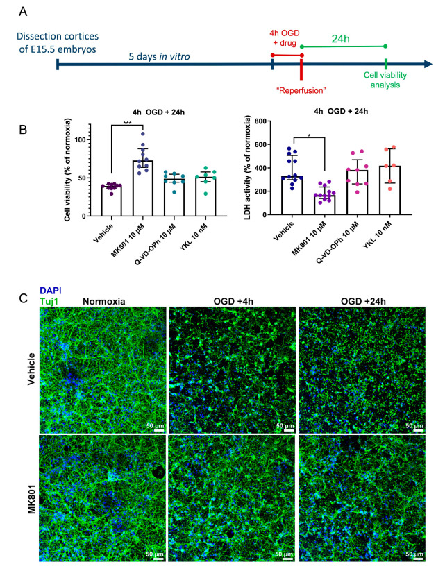
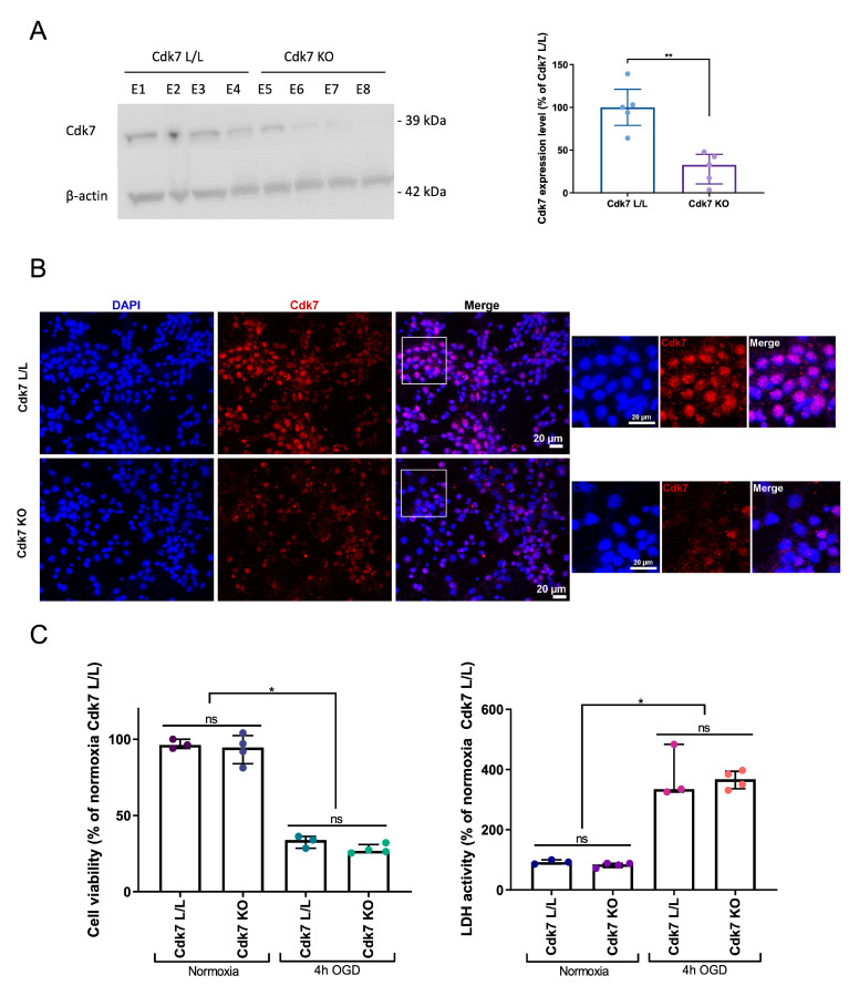
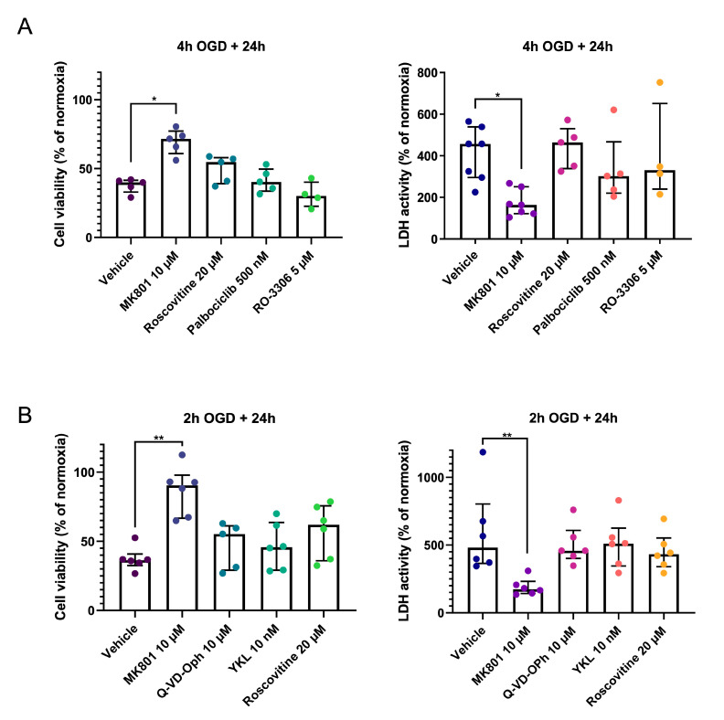
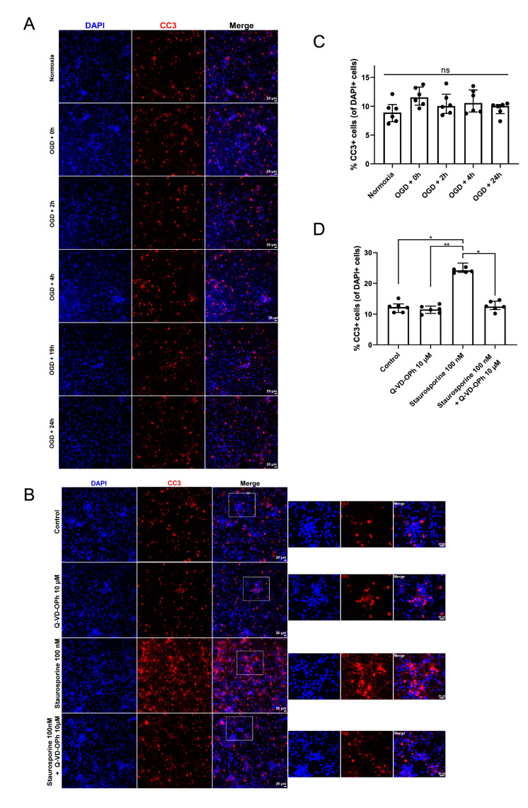
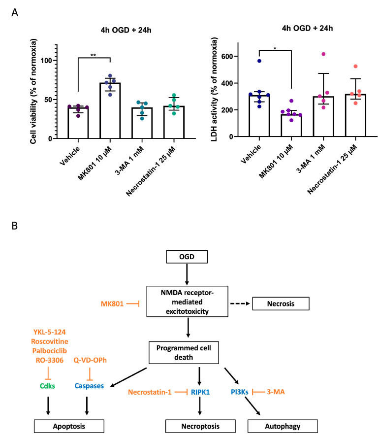
Previous research has shown that cyclin-dependent kinases (Cdks) that play physiological roles in cell cycle regulation become activated in post-mitotic neurons after ischemic stroke, resulting in apoptotic neuronal death. In this article, we report our results using the widely used oxygen-glucose deprivation (OGD) in vitro model of ischemic stroke on primary mouse cortical neurons to investigate whether Cdk7, as part of the Cdk-activating kinase (CAK) complex that activates cell cycle Cdks, might be a regulator of ischemic neuronal death and may potentially constitute a therapeutic target for neuroprotection. We found no evidence of neuroprotection with either pharmacological or genetic invalidation of Cdk7. Despite the well-established idea that apoptosis contributes to cell death in the ischemic penumbra, we also found no evidence of apoptosis in the OGD model. This could explain the absence of neuroprotection following Cdk7 invalidation in this model. Neurons exposed to OGD seem predisposed to die in an NMDA receptor-dependent manner that could not be prevented further downstream. Given the direct exposure of neurons to anoxia or severe hypoxia, it is questionable how relevant OGD is for modeling the ischemic penumbra. Due to remaining uncertainties about cell death after OGD, caution is warranted when using this in vitro model to identify new stroke therapies.

Keywords: OGD; cell culture; cortex; mouse; neurons.


Conflict of interest statement

The authors declare no conflict of interest.

Figures

Figure 1
Figure 1
Inhibition of the NMDA receptor protects against ischemic neuronal death, but not inhibition of Cdk7 or caspases. (A) Experimental timeline. Primary cortical neurons were obtained from E15.5 mouse embryos. At DIV5, neurons were exposed to 4 h of OGD, while incubated with vehicle (0.1% DMSO) or a specific inhibitor of either Cdk7, the NMDA glutamate receptor, or caspases. Viability and cytotoxicity were assessed 24 h after the end of OGD. (B) Viability of primary cortical neurons exposed to 4 h of OGD in the presence of either vehicle only (0.1% DMSO), the NMDA receptor antagonist MK801, the pan-caspase inhibitor Q-VD-OPh, or the Cdk7 inhibitor YKL-5-124 (“YKL”). Neurons were incubated with 0.1% DMSO in all conditions. Viability was measured with the CCK8 assay (left), while cytotoxicity was quantified using the LDH assay (right). Values are expressed as a percentage of normoxia conditions, measured 24 h after the end of OGD. Each dot represents one biological replicate corresponding to a different pregnant mouse, showing the mean value of 4 replicates (4 separate wells) from one batch of primary cortical neurons. Groups were compared using the Kruskal–Wallis test with Dunn’s multiple comparisons test. Error bars represent the interquartile range. * p ≤ 0.05; *** p ≤ 0.001. (C) Representative immunofluorescence images of primary cortical neurons stained for Tuj1 (green) and DAPI (blue) to show the integrity of neurites after OGD in the presence or absence of the NMDA receptor antagonist MK801. Primary cortical neurons were exposed to normoxia or 4 h of OGD in the presence of either 10 µM of MK801 or vehicle (0.1% DMSO). Cells were fixed at either 4 or 24 h after the end of OGD to investigate neurite integrity.
Figure 2
Figure 2
Genetic deletion of Cdk7 does not improve the survival of primary cortical neurons exposed to OGD. (A) Western blot showing Cdk7 levels in primary cortical cultures obtained from either Cdk7lox/lox Nex+/+ (“Cdk7 L/L”; embryo (E) 1-4) or Cdk7lox/lox NexCre/+ (“Cdk7 KO”; E5-8) mice. Western blots were quantified using FIJI/ImageJ and normalized to levels of the housekeeping gene β-actin. Protein levels are expressed as % of the mean of the “Cdk7 L/L” condition for that gel. Each dot on the histogram represents a protein band, corresponding to one batch of cortical cells obtained from one embryo. Statistical significance was investigated using the Mann–Whitney test. (B) Representative immunofluorescence staining for Cdk7 (red) and counterstaining with DAPI (blue) in primary cortical neurons obtained from either Cdk7lox/lox Nex+/+ (“Cdk7 L/L”) or Cdk7lox/lox NexCre/+ (“Cdk7 KO”) mice. Cdk7 is present in virtually all cells obtained from Cdk7lox/lox mice, while its expression is markedly decreased in the KO condition. (C) Viability (CCK8 assay) and cytotoxicity (LDH assay) of primary cortical neurons obtained from either Cdk7lox/lox Nex+/+ (“Cdk7 L/L”) or Cdk7lox/lox NexCre/+ (“Cdk7 KO”) mice were measured 24 h after OGD. Each dot represents the mean value of 4 replicates for one embryo. Groups were compared pairwise using a Kruskal–Wallis test with Dunn’s multiple comparisons test. Error bars: interquartile range. * p ≤ 0.05; ** p ≤ 0.01; ns = not statistically significant.
Figure 3
Figure 3
Pharmacological inhibition of Cdk7 targets does not improve neuronal survival after IRI. Results are not changed by shortening the duration of OGD. (A) Viability (CCK8 assay) and cytotoxicity (LDH assay) of primary cortical neurons exposed to 4 h of OGD in the presence of either vehicle only (0.1% DMSO), positive control MK801, pan-Cdk inhibitor roscovitine, Cdk4/6 inhibitor Palbociclib, or Cdk1/Cdk2 inhibitor RO-3306. Each dot represents the mean of 4 replicates for one batch of primary cortical neurons (each batch is obtained from a separate pregnant mouse). (B) Viability (CCK8 assay) and cytotoxicity (LDH assay) of primary cortical neurons exposed to 2 h of OGD in the presence of either vehicle only (0.1% DMSO), positive control MK801, pan-caspase inhibitor Q-VD-OPh, Cdk7 inhibitor YKL-5-124 (“YKL”), or the broad Cdk inhibitor roscovitine. Each dot represents the mean of 4 replicates for one batch of primary cortical neurons (from a separate pregnant mouse). For all experiments, groups were compared using the Kruskal–Wallis test with Dunn’s multiple comparisons test. Error bars: interquartile range. * p ≤ 0.05; ** p ≤ 0.01.
Figure 4
Figure 4
OGD does not result in marked apoptosis of primary cortical neurons. (A) Representative immunofluorescence images for cleaved caspase-3 (CC3; red) expression as a readout of apoptosis in primary cortical neurons at several time points after OGD (0–24 h). (B) Representative immunofluorescence staining for CC3 expression (red) in primary cortical neurons incubated either with vehicle (0.1% DMSO), 10 µM of the caspase inhibitor Q-VD-OPh, 100 nM of staurosporine, or with both staurosporine and Q-VD-OPh. (C,D) The number of CC3+ cells was counted using FIJI/ImageJ and expressed as a percentage of the total number of cells, determined using DAPI counterstaining (blue). Each dot on the histogram represents the average from 4 different fields of view for one coverslip. Statistical analyses were performed using the Kruskal–Wallis test with Dunn’s multiple comparisons test. Error bars: interquartile range. * p ≤ 0.05; ** p ≤ 0.01; ns = not statistically significant.
Figure 5
Figure 5
Pharmacological inhibition of autophagy and necroptosis does not prevent neurons from dying in response to OGD. (A) Viability of primary cortical neurons exposed to 4 h of OGD in the presence of either vehicle only (0.1% DMSO), MK801 (an NMDA receptor antagonist), 3-MA (an autophagy inhibitor), or necrostatin-1 (an inhibitor of necroptosis). Viability was measured with the CCK8 assay (left), while cytotoxicity was quantified using the LDH assay (right). Values are expressed as a percentage of the normoxia condition, measured 24 h after the end of OGD. Each dot represents the mean value of 4 replicates for a unique batch of primary cortical neurons (each batch is obtained from a separate pregnant mouse). Groups were compared using Kruskal–Wallis test with Dunn’s multiple comparisons test. Error bars: interquartile range. * p ≤ 0.05; ** p ≤ 0.01. (B) Scheme of the hypothetic pathways involved in neuronal cell death following OGD.

Similar articles

Cited by

References

    1. Deb P., Sharma S., Hassan K.M. Pathophysiologic mechanisms of acute ischemic stroke: An overview with emphasis on therapeutic significance beyond thrombolysis. Pathophysiology. 2010;17:197–218. doi: 10.1016/j.pathophys.2009.12.001. - DOI - PubMed
    1. Maida C.D., Norrito R.L., Daidone M., Tuttolomondo A., Pinto A. Neuroinflammatory Mechanisms in Ischemic Stroke: Focus on Cardioembolic Stroke, Background, and Therapeutic Approaches. Int. J. Mol. Sci. 2020;21:6454. doi: 10.3390/ijms21186454. - DOI - PMC - PubMed
    1. Kroemer G., Galluzzi L., Vandenabeele P., Abrams J., Alnemri E.S., Baehrecke E.H., Blagosklonny M.V., El-Deiry W.S., Golstein P., Green D.R., et al. Classification of cell death: Recommendations of the Nomenclature Committee on Cell Death 2009. Cell Death Differ. 2009;16:3–11. doi: 10.1038/cdd.2008.150. - DOI - PMC - PubMed
    1. Nikoletopoulou V., Markaki M., Palikaras K., Tavernarakis N. Crosstalk between apoptosis, necrosis and autophagy. Biochim. Biophys. Acta. 2013;1833:3448–3459. doi: 10.1016/j.bbamcr.2013.06.001. - DOI - PubMed
    1. Tuo Q.Z., Zhang S.T., Lei P. Mechanisms of neuronal cell death in ischemic stroke and their therapeutic implications. Med. Res. Rev. 2022;42:259–305. doi: 10.1002/med.21817. - DOI - PubMed