. 2023 Apr 11;24(8):7072.
doi: 10.3390/ijms24087072.

Lipid-Sensing Receptor FFAR4 Modulates Pulmonary Epithelial Homeostasis following Immunogenic Exposures Independently of the FFAR4 Ligand Docosahexaenoic Acid (DHA)

Affiliations

Lipid-Sensing Receptor FFAR4 Modulates Pulmonary Epithelial Homeostasis following Immunogenic Exposures Independently of the FFAR4 Ligand Docosahexaenoic Acid (DHA)

Stefanie N Sveiven et al. Int J Mol Sci. .

Abstract

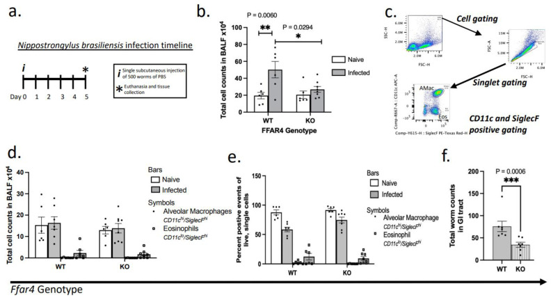
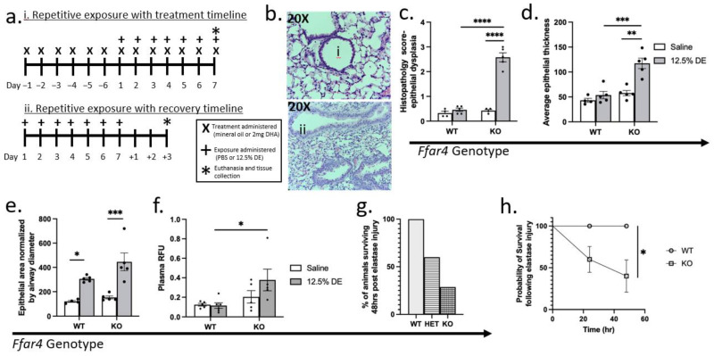
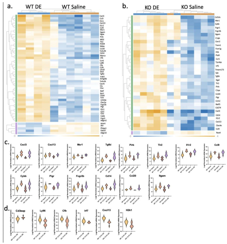
The role of pulmonary free fatty acid receptor 4 (FFAR4) is not fully elucidated and we aimed to clarify the impact of FFAR4 on the pulmonary immune response and return to homeostasis. We employed a known high-risk human pulmonary immunogenic exposure to extracts of dust from swine confinement facilities (DE). WT and Ffar4-null mice were repetitively exposed to DE via intranasal instillation and supplemented with docosahexaenoic acid (DHA) by oral gavage. We sought to understand if previous findings of DHA-mediated attenuation of the DE-induced inflammatory response are FFAR4-dependent. We identified that DHA mediates anti-inflammatory effects independent of FFAR4 expression, and that DE-exposed mice lacking FFAR4 had reduced immune cells in the airways, epithelial dysplasia, and impaired pulmonary barrier integrity. Analysis of transcripts using an immunology gene expression panel revealed a role for FFAR4 in lungs related to innate immune initiation of inflammation, cytoprotection, and immune cell migration. Ultimately, the presence of FFAR4 in the lung may regulate cell survival and repair following immune injury, suggestive of potential therapeutic directions for pulmonary disease.

Keywords: DHA; FFAR4; GPR120; exposure; inflammation; lung.


Conflict of interest statement

The authors declare no conflict of interest.

Figures

Figure 1
Figure 1
Significantly dampened immune recruitment in Ffar4−/− following repetitive pulmonary dust exposure with no changes to cell populations. (a) A schematic of the experimental timeline of repetitive exposure with treatment. Mice were given a 6-day pre-treatment of mineral oil or DHA by oral gavage, followed by a 7-day concurrent treatment with exposure to saline or DE. Mice were euthanized 5 h after the final doses on day 7 of the exposure period. (b) Representative DiffQuick-stained cytospins of BAL cells for saline- and DE-exposed mice show macrophages (m) and neutrophils (n) in the airways (20× and 40×). Repetitive DE exposure over 7 days significantly (three-way ANOVA followed by Tukey’s post hoc test) induces BALF cell influx in an exposure- and genotype-dependent manner (c,d). This influx is marked by neutrophilia in DiffQuick-stained-cytospins determined by cell differential analysis from masked samples (e). There were no significant changes in cell differentials between genotypes, though exposure induced neutrophilia (main effect of exposure: p < 0.0001). (f) H & E stained FFPE lung sections were masked and scored for vascular inflammation. Inflammation was induced by DE exposure in both WT and KO compared with their genotype-matched controls (Adj. p = 0.0127 and <0.0001 respectively). Ffar4 KO mice had significantly higher inflammatory scores than WT (Adj. p = 0.0095). Experiments were performed in triplicate (at minimum) with all groups represented in each separate experiment. * p < 0.05; ** p < 0.01; **** p < 0.0001.
Figure 2
Figure 2
The Ffar4−/− phenotype of dampened immune influx without changes to cell populations is confirmed in another model: Nippostrongylus brasiliensis (Nb) infection. (a) A schematic of the helminth infection timeline. Mice were subcutaneously injected with 500 Nippostrongylus brasiliensis L3 larvae in 200 μL PBS or PBS only on day 0. Mice were euthanized on day 5 post-infection. (b) Helminth-infected WT mice had significantly elevated cell counts in the airway compared with their naïve controls, as measured by total cell counts in the BALF; this effect was significantly dampened by Ffar4-deficiency, being comparable to naïve controls. (ce) BAL cell differentials, determined by flow cytometry, were not significantly altered by Ffar4 presence, though the infection significantly impacted cell differentials by promoting eosinophilia in the airways (two-way ANOVA, main effect p < 0.0001). Expressing the data as totals or percentages did not alter this outcome. These data were generated from two separate experiments with representative groups included in each experiment. (f) Ffar4-deficiency dampened the presence of worms in the GI tract, with significantly fewer worms counted from the jejunum of Ffar4-null mice. * p < 0.05; ** p < 0.01; *** p < 0.001.
Figure 3
Figure 3
Lack of Ffar4 impairs airway epithelial barrier homeostasis. (a) Schematic of repetitive exposure timeline without (i) and with (ii) the recovery period. The recovery studies did not include treatments, and mice were left to recover without exposure for three days. On the third day of recovery, 1 h before euthanizing, mice were given intranasal RhoB-dextran in PBS. X’s represent oral gavage treatments with mineral oil (vehicle control) or 2 mg DHA, plus signs (+) represent the administration of intranasal exposure (PBS or 12.5% DE), and the asterisk (*) signifies euthanasia. If euthanasia fell on the day of an exposure, animals were euthanized 5 h after administration. (b) H & E-stained FFPE lung sections reveal a dysplastic epithelial appearance (representative images of (i) normal and (ii) dysplastic epithelium at 20×). Ffar4-deficient mice exposed to DE had significantly higher airway epithelial dysplasia scores compared with DE-exposed WT and naïve KO mice ((c), Adj. p < 0.0001 for each), and similar findings were observed in quantified epithelial thickness ((d), Adj. p = 0.0001 and 0.0002, respectively). Airway area normalized to diameter was only significant in both dust-exposed WT and KO mice compared with their respective genotype-matched saline controls ((e), Adj. p = 0.0285 and 0.004 respectively). (f) Using intranasal Rhodamine B dextran delivery, Ffar4-deficient mice had significantly greater plasma fluorescence following repetitive instillations of DE plus three days for repair (Adj. p = 0.0081). (g,h) Ffar4-deficient mice had significantly reduced survival in an acute epithelial injury model of a single dose of intranasal porcine pancreatic elastase ((h), Log-rank test, p = 0.0224). ** p < 0.01; **** p < 0.001; **** p < 0.0001.
Figure 4
Figure 4
Lack of Ffar4 and reduced YAP signal in lung immunofluorescence. Immunofluorescence was performed on FFPE lung sections from mice repetitively exposed to 12.5% DE or saline (5 μm sections imaged at 20×; scale bars are 200 μm). (a) Anti-murine YAP or TAZ antibodies were used, followed by secondary antibody incubation, then counter-stained with DAPI. (b) YAP intensity showed a significant main-effect of genotype (2-way ANOVA, p = 0.0424), while TAZ was not significant in these comparisons.
Figure 5
Figure 5
Dust-induced differentially expressed genes in total lung RNA from repetitively exposed mice. Total lung bulk RNA from mice repetitively exposed to 12.5% DE or saline was analyzed by the mouse immunology NanoString nCounter panel. Significant genes (Adj. p ≤ 0.05) with a fold-change of +/−1.5× are reported as heat maps of differentially expressed genes (DEG) in DE-exposed WT mice compared with WT saline controls ((a), 51 genes) or DE-exposed KO mice compared with KO saline controls ((b), 34 genes). One of the KO DE samples clusters with the saline group, which could be an artifact of poor intranasal delivery. Violin plots of the (c) 14 DEGs overlapping in both DE-exposed WT and KO samples compared with their genotype-matched saline controls (Adj. p < 0.05, FC: +/−1.5×) and (d) 5 DEGs between DE-exposed KO compared with WT (p < 0.05, FC: +/−1.5×).

Similar articles

Cited by

References

    1. Nordgren T.M., Friemel T.D., Heires A.J., Poole J.A., Wyatt T.A., Romberger D.J. The omega-3 fatty acid docosahexaenoic acid attenuates organic dust-induced airway inflammation. Nutrients. 2014;6:5434–5452. doi: 10.3390/nu6125434. - DOI - PMC - PubMed
    1. Nordgren T.M., Heires A.J., Bailey K.L., Katafiasz D.M., Toews M.L., Wichman C.S., Romberger D.J. Docosahexaenoic acid enhances amphiregulin-mediated bronchial epithelial cell repair processes following organic dust exposure. Am. J. Physiol. Lung Cell Mol. Physiol. 2018;314:L421–L431. doi: 10.1152/ajplung.00273.2017. - DOI - PMC - PubMed
    1. Oh D.Y., Walenta E. Omega-3 Fatty Acids and FFAR4. Front. Endocrinol. 2014;5:115. doi: 10.3389/fendo.2014.00115. - DOI - PMC - PubMed
    1. Mone P., Varzideh F., Kansakar U., Infante C., Lombardi A., de Donato A., Frullone S., Santulli G. Omega-3 fatty acids coordinate glucose and lipid metabolism in diabetic patients. Lipids Health Dis. 2022;21:31. doi: 10.1186/s12944-022-01642-w. - DOI - PMC - PubMed
    1. Skulas-Ray A.C., Wilson P.W.F., Harris W.S., Brinton E.A., Kris-Etherton P.M., Richter C.K., Jacobson T.A., Engler M.B., Miller M., Robinson J.G., et al. Omega-3 Fatty Acids for the Management of Hypertriglyceridemia: A Science Advisory From the American Heart Association. Circulation. 2019;140:e673–e691. doi: 10.1161/CIR.0000000000000709. - DOI - PubMed