

Dynamics of the Equine Placental DNA Methylome and Transcriptome from Mid- to Late Gestation
- PMID: 37108254
- PMCID: PMC10139181
- DOI: 10.3390/ijms24087084
Dynamics of the Equine Placental DNA Methylome and Transcriptome from Mid- to Late Gestation
Abstract
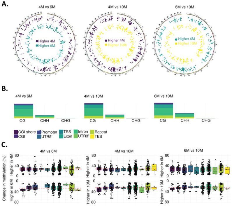
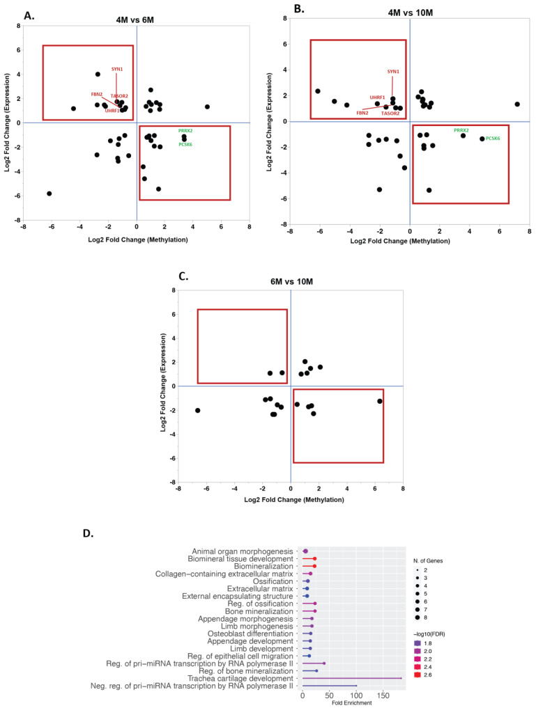
The placenta is a temporary organ that is essential for the survival of the fetus, with a lifelong effect on the health of both the offspring and the dam. The functions of the placenta are controlled by its dynamic gene expression during gestation. In this study, we aimed to investigate the equine placental DNA methylome as one of the fundamental mechanisms that controls the gene expression dynamic. Chorioallantois samples from four (4M), six (6M), and ten (10M) months of gestation were used to map the methylation pattern of the placenta. Globally, methylation levels increased toward the end of gestation. We identified 921 differentially methylated regions (DMRs) between 4M and 6M, 1225 DMRs between 4M and 10M, and 1026 DMRs between 6M and 10M. A total of 817 genes carried DMRs comparing 4M and 6M, 978 comparing 4M and 10M, and 804 comparing 6M and 10M. We compared the transcriptomes between the samples and found 1381 differentially expressed genes (DEGs) when comparing 4M and 6M, 1428 DEGs between 4M and 10M, and 741 DEGs between 6M and 10M. Finally, we overlapped the DEGs and genes carrying DMRs (DMRs-DEGs). Genes exhibiting (a) higher expression, low methylation and (b) low expression, high methylation at different time points were identified. The majority of these DMRs-DEGs were located in introns (48.4%), promoters (25.8%), and exons (17.7%) and were involved in changes in the extracellular matrix; regulation of epithelial cell migration; vascularization; and regulation of minerals, glucose, and metabolites, among other factors. Overall, this is the first report highlighting the dynamics in the equine placenta methylome during normal pregnancy. The findings presented serve as a foundation for future studies on the impact of abnormal methylation on the outcomes of equine pregnancies.
Keywords: chorioallantois; differentially methylated regions; horse; methylation; placental methylome; reduced representation bisulfate sequencing.
Conflict of interest statement
The authors declare no conflict of interest.
Figures


Similar articles
-
Sex-based disparities in DNA methylation and gene expression in late-gestation mouse placentas.Biol Sex Differ. 2024 Jan 6;15(1):2. doi: 10.1186/s13293-023-00577-w. Biol Sex Differ. 2024. PMID: 38183126 Free PMC article.
-
Integrative analysis of methylomic and transcriptomic data in fetal sheep muscle tissues in response to maternal diet during pregnancy.BMC Genomics. 2018 Feb 6;19(1):123. doi: 10.1186/s12864-018-4509-0. BMC Genomics. 2018. PMID: 29409445 Free PMC article.
-
Methylome and transcriptome data integration reveals aberrantly regulated genes in equine sarcoids.Biochimie. 2023 Oct;213:100-113. doi: 10.1016/j.biochi.2023.05.008. Epub 2023 May 19. Biochimie. 2023. PMID: 37211255
-
Placental DNA methylation profile as predicting marker for autism spectrum disorder (ASD).Mol Med. 2023 Jan 16;29(1):8. doi: 10.1186/s10020-022-00593-3. Mol Med. 2023. PMID: 36647002 Free PMC article. Review.
-
Recent progress towards understanding the role of DNA methylation in human placental development.Reproduction. 2016 Jul;152(1):R23-30. doi: 10.1530/REP-16-0014. Epub 2016 Mar 29. Reproduction. 2016. PMID: 27026712 Free PMC article. Review.
Cited by
-
In search of epigenetic hallmarks of different tissues: an integrative omics study of horse liver, lung, and heart.Mamm Genome. 2024 Dec;35(4):600-620. doi: 10.1007/s00335-024-10057-0. Epub 2024 Aug 14. Mamm Genome. 2024. PMID: 39143382 Free PMC article.
References
-
- Novakovic B., Yuen R.K., Gordon L., Penaherrera M.S., Sharkey A., Moffett A., Craig J.M., Robinson W.P., Saffery R. Evidence for widespread changes in promoter methylation profile in human placenta in response to increasing gestational age and environmental/stochastic factors. BMC Genom. 2011;12:529. doi: 10.1186/1471-2164-12-529. - DOI - PMC - PubMed
