

Membrane Lesions and Reduced Life Span of Red Blood Cells in Preeclampsia as Evidenced by Atomic Force Microscopy
- PMID: 37108270
- PMCID: PMC10138579
- DOI: 10.3390/ijms24087100
Membrane Lesions and Reduced Life Span of Red Blood Cells in Preeclampsia as Evidenced by Atomic Force Microscopy
Abstract
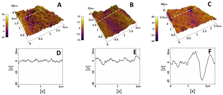
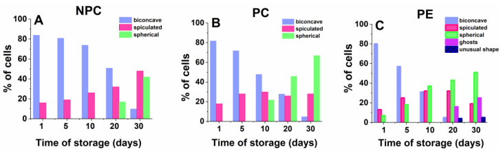
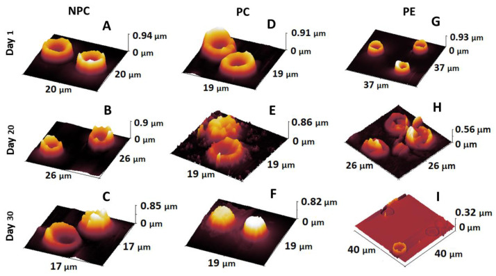
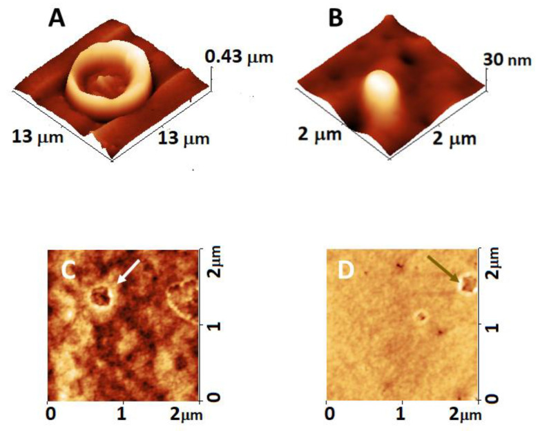
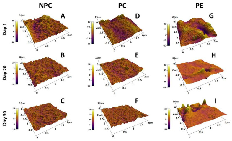
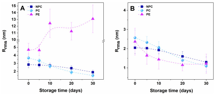
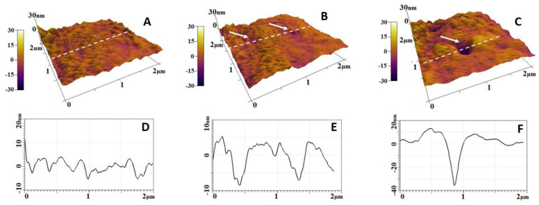
Preeclampsia (PE) presents with maternal de novo hypertension and significant proteinuria and is one of the leading causes of maternal and perinatal morbidity and mortality with unknown etiology. The disease is associated with inflammatory vascular response and severe red blood cell (RBC) morphology changes. This study examined the nanoscopic morphological changes of RBCs from PE women versus normotensive healthy pregnant controls (PCs) and non-pregnant controls (NPCs) applying atomic force microscopy (AFM) imaging. The results revealed that the membrane of fresh PE RBCs differed significantly from healthy ones by the presence of invaginations and protrusions and an increased roughness value (Rrms) (4.7 ± 0.8 nm for PE vs. 3.8 ± 0.5 nm and 2.9 ± 0.4 nm for PCs and NPCs, respectively). PE-cells aging resulted in more pronounced protrusions and concavities, with exponentially increasing Rrms values, in contrast to the controls, where the Rrms parameter decreased linearly with time. The Rrms, evaluated on a 2 × 2 µm2 scanned area, for senescent PE cells (13 ± 2.0 nm) was significantly higher (p < 0.01) than that of PCs (1.5 ± 0.2 nm) and NPCs (1.9 ± 0.2 nm). Furthermore, the RBCs from PE patients appeared fragile, and often only ghosts were observed instead of intact cells at 20-30 days of aging. Oxidative-stress simulation on healthy cells led to RBC membrane features similar to those observed for PE cells. The results demonstrate that the most pronounced effects on RBCs in PE patients are related to impaired membrane homogeneity and strongly altered roughness values, as well as to vesiculation and ghost formation in the course of cell aging.
Keywords: atomic force microscopy; cell senescence; membrane impairment; membrane roughness; preeclampsia; red blood cells.
Conflict of interest statement
The authors declare no potential conflict of interest.
Figures
Similar articles
-
Assessment of Red Blood Cell Aggregation in Preeclampsia by Microfluidic Image Flow Analysis-Impact of Oxidative Stress on Disease Severity.Int J Mol Sci. 2024 Mar 27;25(7):3732. doi: 10.3390/ijms25073732. Int J Mol Sci. 2024. PMID: 38612543 Free PMC article.
-
The how, when, and why of the aging signals appearing on the human erythrocyte membrane: an atomic force microscopy study of surface roughness.Nanomedicine. 2010 Dec;6(6):760-8. doi: 10.1016/j.nano.2010.06.004. Epub 2010 Jul 30. Nanomedicine. 2010. PMID: 20603227
-
Morphological changes induced in erythrocyte by amyloid beta peptide and glucose depletion: A combined atomic force microscopy and biochemical study.Biochim Biophys Acta Biomembr. 2019 Jan;1861(1):236-244. doi: 10.1016/j.bbamem.2018.07.009. Epub 2018 Jul 21. Biochim Biophys Acta Biomembr. 2019. PMID: 30040926
-
Nanoscale Surface Characterization of Human Erythrocytes by Atomic Force Microscopy: A Critical Review.IEEE Trans Nanobioscience. 2015 Sep;14(6):625-33. doi: 10.1109/TNB.2015.2424674. Epub 2015 Apr 28. IEEE Trans Nanobioscience. 2015. PMID: 25935044 Review.
-
Structure and function in native and pathological erythrocytes: a quantitative view from the nanoscale.Micron. 2012 Dec;43(12):1273-86. doi: 10.1016/j.micron.2012.03.019. Epub 2012 Apr 3. Micron. 2012. PMID: 22537716 Review.
Cited by
-
Red Blood Cell Storage with Xenon: Safe or Disruption?Cells. 2024 Feb 27;13(5):411. doi: 10.3390/cells13050411. Cells. 2024. PMID: 38474375 Free PMC article.
-
Assessment of Red Blood Cell Aggregation in Preeclampsia by Microfluidic Image Flow Analysis-Impact of Oxidative Stress on Disease Severity.Int J Mol Sci. 2024 Mar 27;25(7):3732. doi: 10.3390/ijms25073732. Int J Mol Sci. 2024. PMID: 38612543 Free PMC article.
References
-
- Shaikh N., Nahid S., Ummunnisa F., Fatima I., Hilani M., Gul A., Basha A.A., Yahia W., Hail F.A., Elfil H., et al. Preeclampsia: From Etiopathology to Organ Dysfunction. In: Abduljabbar H., editor. Preeclampsia. IntechOpen; Jeddah, Saudi Arabia: 2021.
MeSH terms
Grants and funding
LinkOut - more resources
Full Text Sources
Miscellaneous
