. 2023 Apr 12;24(8):7117.
doi: 10.3390/ijms24087117.

SM22α Deletion Contributes to Neurocognitive Impairment in Mice through Modulating Vascular Smooth Muscle Cell Phenotypes

Affiliations

SM22α Deletion Contributes to Neurocognitive Impairment in Mice through Modulating Vascular Smooth Muscle Cell Phenotypes

Xin Xu et al. Int J Mol Sci. .

Abstract

Considerable evidence now indicates that cognitive impairment is primarily a vascular disorder. The depletion of smooth muscle 22 alpha (SM22α) contributes to vascular smooth muscle cells (VSMCs) switching from contractile to synthetic and proinflammatory phenotypes in the context of inflammation. However, the role of VSMCs in the pathogenesis of cognitive impairment remains undetermined. Herein, we showed a possible link between VSMC phenotypic switching and neurodegenerative diseases via the integration of multi-omics data. SM22α knockout (Sm22α-/-) mice exhibited obvious cognitive impairment and cerebral pathological changes, which were visibly ameliorated by the administration of AAV-SM22α. Finally, we confirmed that SM22α disruption promotes the expression of SRY-related HMG-box gene 10 (Sox10) in VSMCs, thereby aggravating the systemic vascular inflammatory response and ultimately leading to cognitive impairment in the brain. Therefore, this study supports the idea of VSMCs and SM22α as promising therapeutic targets in cognitive impairment to improve memory and cognitive decline.

Keywords: SRY-related HMG-box gene 10; cognitive impairment; inflammation; smooth muscle 22-alpha; vascular smooth muscle cells.


Conflict of interest statement

The authors declare that they have no competing interests.

Figures

Figure 1
Figure 1
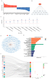
Multi-omics points to a possible link between VSMC phenotypic switching and neurodegenerative diseases. (A) KEGG pathway enrichment of differentially expressed proteins between contractile and synthetic VSMCs in mice from proteomic analyses (n = 3 independent samples). (B) Heatmap of each lipid class between contractile and synthetic VSMCs in mice from lipidomic analyses (n = 4 independent samples). (C) KEGG pathway enrichment of upregulated differentially expressed mRNAs in the arteries of Sm22α−/− mice compared with WT mice from transcriptomic analyses (n = 8 mice per group).
Figure 2
Figure 2
Depletion of SM22α leads to pathological changes in the hippocampus region of the mouse brain. (A) Representative images of HE staining in the hippocampus of the WT and Sm22α−/− mice. (B) Representative images of Nissl staining in the hippocampus of the WT and Sm22α−/− mice are shown on the left, with the statistical results regarding the number of neurons on the right. CA1, hippocampal CA1 layer; DG, dentate gyrus. ** p < 0.01.
Figure 3
Figure 3
The Sm22α−/− mice exhibited cognitive impairment. (A) Schematics of the experimental design for the cognitive behavioral study. OFT, open field test; NOR, novel object recognition test; MWN, Morris water maze test; D, day. (BD) Performances in the OFT were recorded for 5 min. (B) Distance traveled in the center. (C) Time spent in the center. (D) Total travel distance. (E) Discrimination ratio of novel versus familiar objects during NOR. (FL) Performances in the MWM during the training days and probe test. (F) Escape latency to the platform during the training days. (G) Latency to first reaching the platform and (H) representative track images of the mice in the probe test with the platform. (I) Distance in the target quadrant, (J) time spent in the target quadrant, (K) entry number in the platform zone, and (L) representative track images of the mice during the 60 s probe test without the platform. Data are represented as the mean ± SD (n = 8 mice per group). * p < 0.05, ** p < 0.01, and *** p < 0.001.
Figure 4
Figure 4
Administration of AAV-SM22α improved the cognitive impairment in the Sm22α−/− mice. (AC) Performance in the OFT was recorded for 5 min. (A) Distance traveled in the center. (B) Time spent in the center. (C) Total travel distance. (D) Discrimination ratio of novel versus familiar objects during the NOR. (EK) Performance in the MWM in the training days and probe test. (E) Escape latency to the platform during the training days. (F) Latency to first reach the platform and (G) representative track images of the mice in the probe test with the platform. (H) Distance in the target quadrant, (I) time spent in the target quadrant, (J) entry number in the platform zone, and (K) representative track images of the mice during the 60 s probe test without the platform. Data are represented as the mean ± SD (n = 8 mice per group). * p < 0.05, ** p < 0.01, and *** p < 0.001.
Figure 5
Figure 5
Sox10 is closely associated with neurodegenerative diseases. (A) Top differentially expressed genes in modulated VSMCs of the neointima of those mice with ligated carotid arteries from single-cell RNA sequencing (n = 30 mice). (B) Potential transcriptional regulatory target genes of Sox10 in modulated VSMCs. (C) Differential expression of Sox10 between AD cases and controls across brain regions from the AMP-AD knowledge portal. The box plot depicts how the differential expression of Sox10 (purple dot) compares to the expression of other genes in a given tissue. Meaningful differential expression is considered to be a log2 fold change value greater than 0.263 or less than –0.263 (red line). (D) The interaction network of transcription factor Sox10 and its downstream target genes detected in the mouse brain using the GRNdb online network (http://www.grndb.com/; accessed on 6 November 2022). (E) Gene ontology (GO) terms of the Sox10 downstream target genes in the mouse brain. BP, biological process; CC, cellular component; MF, molecular function. (F) Sankey plot of the pathways enriched for the Sox10 downstream target genes in the mouse brain. Genes associated with neurodegenerative diseases are shown in red.
Figure 6
Figure 6
Sox10 expression is associated with the inflammatory response in the mouse brain. (A) Representative images of Sox10 immunohistochemical staining in the hippocampus of the WT and Sm22α−/− mice are shown on the left, with the corresponding statistical result on the right. (B) Representative Western blots and quantitative analysis of the brain in the WT and Sm22α−/− mice treated with PDGF-BB ex vivo. (C) Representative Western blots and quantitative analysis of the aorta in the WT and Sm22α−/− mice treated with PDGF-BB ex vivo. (D) Representative Western blots and quantitative analysis of VSMCs treated with PDGF-BB at different time points. (E) Representative Western blots and quantitative analysis of Sox10 in HEK 293A cells treated with TNF-α after pretreatment with CHX or MG132 for the indicated times. (F) Sequence alignment of Sox10 Ser24 from different species. (G) Representative Western blots and quantitative analysis of Sox10 in HEK 293A cells transfected with Flag-Sox10-WT or Flag-Sox10-S24A, and then treated with TNF-α. (H) Representative Western blots and quantitative analysis of Sox10 in HEK 293A cells transfected with Flag-Sox10-WT or Flag-Sox10-S24A, and then pretreatment with or without MG132 before adding TNF-α. Data are represented as the mean ± SD. * p < 0.05, ** p < 0.01, and *** p < 0.001.

Similar articles

Cited by

References

    1. Cheng Y.W., Chen T.F., Chiu M.J. From mild cognitive impairment to subjective cognitive decline: Conceptual and methodological evolution. Neuropsychiatr. Dis. Treat. 2017;13:491–498. doi: 10.2147/NDT.S123428. - DOI - PMC - PubMed
    1. Livingston G., Huntley J., Sommerlad A., Ames D., Ballard C., Banerjee S., Brayne C., Burns A., Cohen-Mansfield J., Cooper C., et al. Dementia prevention, intervention, and care: 2020 report of the Lancet Commission. Lancet. 2020;396:413–446. - PMC - PubMed
    1. Tian Z., Ji X., Liu J. Neuroinflammation in Vascular Cognitive Impairment and Dementia: Current Evidence, Advances, and Prospects. Int. J. Mol. Sci. 2022;23:6224. doi: 10.3390/ijms23116224. - DOI - PMC - PubMed
    1. van der Flier W.M., Skoog I., Schneider J.A., Pantoni L., Mok V., Chen C.L.H., Scheltens P. Vascular cognitive impairment. Nat. Rev. Dis. Prim. 2018;4:18003. doi: 10.1038/nrdp.2018.3. - DOI - PubMed
    1. Azarpazhooh M.R., Avan A., Cipriano L.E., Munoz D.G., Sposato L.A., Hachinski V. Concomitant vascular and neurodegenerative pathologies double the risk of dementia. Alzheimers Dement. 2018;14:148–156. doi: 10.1016/j.jalz.2017.07.755. - DOI - PubMed

LinkOut - more resources