. 2023 Apr 13;24(8):7175.
doi: 10.3390/ijms24087175.

Assessing the Orthogonality of Phage-Encoded RNA Polymerases for Tailored Synthetic Biology Applications in Pseudomonas Species

Affiliations

Assessing the Orthogonality of Phage-Encoded RNA Polymerases for Tailored Synthetic Biology Applications in Pseudomonas Species

Eveline-Marie Lammens et al. Int J Mol Sci. .

Abstract

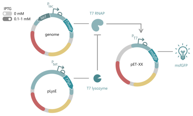
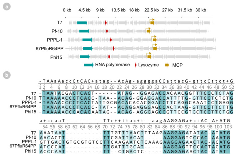
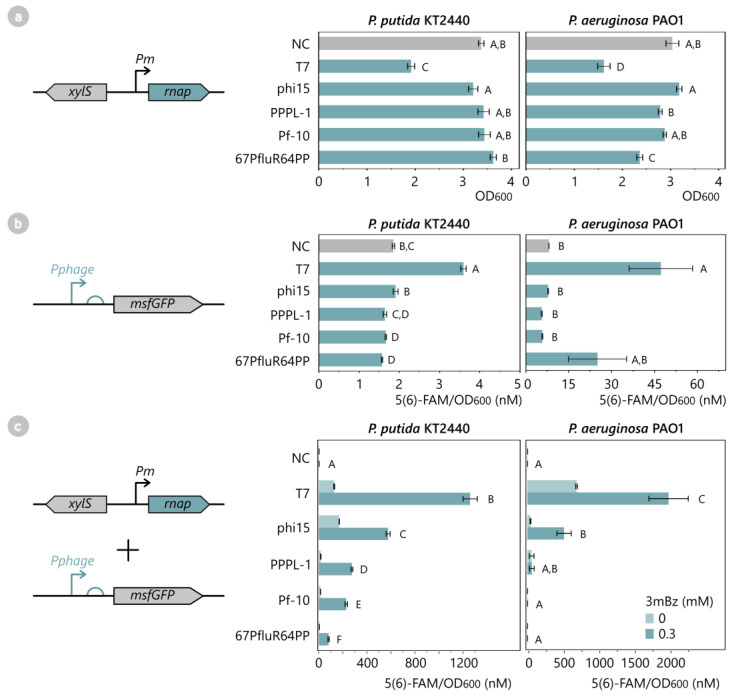
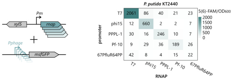
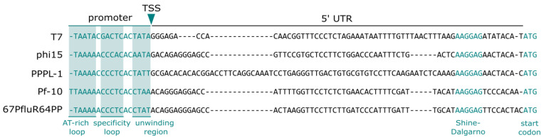
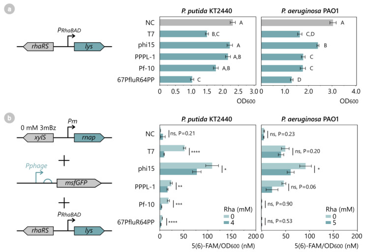
The phage T7 RNA polymerase (RNAP) and lysozyme form the basis of the widely used pET expression system for recombinant expression in the biotechnology field and as a tool in microbial synthetic biology. Attempts to transfer this genetic circuitry from Escherichia coli to non-model bacterial organisms with high potential have been restricted by the cytotoxicity of the T7 RNAP in the receiving hosts. We here explore the diversity of T7-like RNAPs mined directly from Pseudomonas phages for implementation in Pseudomonas species, thus relying on the co-evolution and natural adaptation of the system towards its host. By screening and characterizing different viral transcription machinery using a vector-based system in P. putida., we identified a set of four non-toxic phage RNAPs from phages phi15, PPPL-1, Pf-10, and 67PfluR64PP, showing a broad activity range and orthogonality to each other and the T7 RNAP. In addition, we confirmed the transcription start sites of their predicted promoters and improved the stringency of the phage RNAP expression systems by introducing and optimizing phage lysozymes for RNAP inhibition. This set of viral RNAPs expands the adaption of T7-inspired circuitry towards Pseudomonas species and highlights the potential of mining tailored genetic parts and tools from phages for their non-model host.

Keywords: Pseudomonas; RNA polymerase; T7-like phages; lysozyme; orthogonality.


Conflict of interest statement

The authors declare no conflict of interest.

Figures

Figure 1
Figure 1
General layout of the T7-based pET system for E. coli. The T7 RNAP is stably integrated into the host genome with an IPTG-inducible expression cassette. In the absence of IPTG (light grey), the system is considered off, while upon addition of IPTG (dark grey, usually 0.1–1 mM IPTG), the T7 RNAP is expressed from the LacI/Plac system. The T7 RNAP drives expression of a gene of interest, here depicted as an msfGFP, from its putative T7 promoter on any pET vector. To express toxic proteins, alternative hosts are available, carrying the pLys vector for expression of the T7 lysozyme, which inhibits transcriptional activity of the T7 RNAP in uninduced conditions. IPTG: isopropyl-β-D-thiogalactopyranoside, T7 RNAP: T7 RNA polymerase, msfGFP: monomeric superfolder green fluorescent protein.
Figure 2
Figure 2
(a) Genomic organization of phages T7, Pf-10, PPPL-1, 67PfluR64PP, and phi15. The RNA polymerase genes, lysozyme genes, and major capsid protein (MCP) genes are indicated in teal, red, and yellow, respectively. (b) Multiple sequence alignment (ClustalOmega) of the predicted phage promoter and 5′ untranslated region (UTR) of the phage’s major capsid protein. Darker shaded background colors indicate a higher level of conservation.
Figure 3
Figure 3
(a) Effect of phage RNAP expression on cell growth of P. putida KT2440 and P. aeruginosa PAO1. The RNAPs of phages T7, phi15, PPPL-1, Pf-10, and 67PfluR64PP were introduced in P. putida and P. aeruginosa, as well as an empty pSTDesX vector as a negative control (NC). All samples were induced with 1 mM 3mBz at OD600 0.3 for 12 h. Bars and error bars represent the mean OD600 and standard error of four biological replicates after 12 h induction, respectively. Samples not connected by the same letters are significantly different (Tukey HSD, α = 0.05). (b) Recognition of the phage promoter by the host RNAP of P. putida KT2440 and P. aeruginosa PAO1. MsfGFP reporter constructs with the phage-specific promoters of T7, phi15, PPPL-1, Pf-10, and 67PfluR64PP and a promoterless construct (NC) were introduced in P. putida or P. aeruginosa and were monitored for 12 h for OD600 and msfGFP levels. As the phage RNAPs were not present, no msfGFP expression was expected unless the phage promoter was also recognized by the host RNAP. Bars and error bars represent the mean value and standard error of four biological replicates after 12 h cell growth, respectively. Samples not connected by the same letters are significantly different (Tukey HSD, α = 0.05). (c) T7-like phage RNAPs generate high msfGFP expression levels from their putative phage promoter in P. putida KT2440 and P. aeruginosa PAO1. All phage RNAPs and corresponding phage promoter-msfGFP reporter constructs, including empty control vectors (NC), were introduced in P. putida and P. aeruginosa and were induced with 0.3 mM 3mBz for a 12 h period. The fluorescent intensity was normalized for OD and expressed as an equivalent 5(6)-FAM concentration (nM). Bars and error bars represent the mean value and standard error of four biological replicates after 12 h induction, respectively. Samples not connected by the same letters are significantly different (Wilcoxon (P. putida) and Student’s t-test (P. aeruginosa), α = 0.05). The complete growth curves and fluorescence expression levels over time of (ac) are provided in Figure S3.
Figure 4
Figure 4
To verify cross-recognition between the phage RNAPs and their promoters, all 25 combinations of RNAPs and phage promoter-msfGFP reporter constructs were introduced in P. putida KT2440 and induced with 0.3 mM 3mBz overnight. The fluorescent intensity was normalized for the OD and expressed as an equivalent 5(6)-FAM concentration (nM). Values represent the mean normalized fluorescence intensity after overnight induction of four biological replicates. The statistical analysis of all pairwise comparisons and a connecting letter report are provided in Tables S4 and S5.
Figure 5
Figure 5
Clustal-omega alignment of the validated promoter and 5′ UTR of the phages’ major capsid protein, separated by the confirmed transcription start site (TSS). Confirmed promoter regions of the T7 promoter and 5′ UTR, namely the AT-rich recognition loop, specificity loop, unwinding region, Shine–Dalgarno sequence, and start codon, are projected on the promoters of the other phages.
Figure 6
Figure 6
(a) Toxicity assay of phage lysozymes in P. putida KT2440 and P. aeruginosa PAO1. All phage lysozymes of T7, phi15, PPPL-1, Pf-10, and 67PfluR64PP, as well as an empty pSTDesR control vector (NC), were introduced in P. putida and P. aeruginosa and were induced with 10 mM Rha at OD600 0.3, after which cell growth was monitored for 12 h. Bars and error bars represent the mean and standard error of four biological replicates. Samples not connected by the same letters are significantly different (Tukey HSD, α = 0.05). (b) Fluorescence assay to analyze the inhibitory effect of the phage lysozyme on its corresponding phage RNAP. All phage RNAPs, lysozymes, and phage promoter-msfGFP reporter constructs of T7, phi15, PPPL-1, Pf-10, and 67PfluR64PP and empty control vectors (NC) were introduced in P. putida and induced with 4–5 mM Rha for 12 h. Data points represent the mean 5(6)-FAM/OD600 value of four biological replicates, while error bars represent the standard error. Significance levels are indicated for one-sided Student’s t-tests, with *: p < 0.05, **: p < 0.01, ***: p < 0.001 and ****: p < 0.0001. Full growth curves and fluorescence expression levels over time of (a,b) are available in Figure S5.
Figure 7
Figure 7
(a) Fluorescence intensity assay to analyze the inhibitory effect of different phage lysozymes on the phi15 RNAP. The phage lysozymes of T7, phi15, PPPL-1, Pf-10, 67PfluR64PP, and an empty pSTDes3 control vector (NC) were introduced in P. putida KT2440 together with the phi15 RNAP and Pphi15-msfGFP reporter construct. All samples were induced with 4 mM Rha at OD600 0.3, after which the fluorescence intensity and cell growth were monitored every half hour for 12 h. Bars and error bars represent the mean 5(6)-FAM/OD600 value and standard error of four biological replicates, respectively. Samples not connected by the same letters are significantly different (Tukey HSD, α = 0.05). (b) Similar fluorescence assay as (a) to assess the inhibitory strength of different phi15 lysozyme mutants on the phi15 RNAP. The wild-type phi15 and Pf-10 lysozymes (WT), as well as phi15 lysozyme mutants (AA1-9 > Pf10(AA1-10)), (G3R), (G3Q), (G3RQ), (K5Q), and (K7N,E8K), were introduced in P. putida, together with the phi15 RNAP and Pphi15-msfGFP reporter construct. All samples were induced with 5 mM Rha for 12 h. Bars and error bars represent the mean 5(6)-FAM/OD600 value and standard error of four biological replicates, respectively. Samples not connected by the same letters are significantly different (Tukey HSD, α = 0.05). (c) Assessment of the inhibitory strength of the phi15 lysozyme (G3RQ) mutant in P. aeruginosa PAO1. The wild-type phi15 lysozyme (WT) and phi15 lysozyme mutant (G3RQ) were introduced in P. aeruginosa, together with the phi15 RNAP and Pphi15-msfGFP reporter construct, and induced with 5 mM Rha for 12 h. Bars and error bars represent the mean 5(6)-FAM/OD600 value and standard error of four biological replicates, respectively. The significance level for a one-sided Student’s t-test is indicated, with **: p < 0.01. Full growth curves and fluorescence expression levels over time of (ac) are available in Figure S9.

Similar articles

Cited by

References

    1. Lammens E.-M., Nikel P.I., Lavigne R. Exploring the Synthetic Biology Potential of Bacteriophages for Engineering Non-Model Bacteria. Nat. Commun. 2020;11:5294. doi: 10.1038/s41467-020-19124-x. - DOI - PMC - PubMed
    1. Davanloo P., Rosenberg A.H., Dunn J.J., Studier F.W. Cloning and Expression of the Gene for Bacteriophage T7 RNA Polymerase. Proc. Natl. Acad. Sci. USA. 1984;81:2035–2039. doi: 10.1073/pnas.81.7.2035. - DOI - PMC - PubMed
    1. Studier F.W. Use of Bacteriophage T7 Lysozyme to Improve an Inducible T7 Expression System. J. Mol. Biol. 1991;219:37–44. doi: 10.1016/0022-2836(91)90855-Z. - DOI - PubMed
    1. Luo R., Lian G., Li H., Han H., Zhou D., Gong X. Ultrasensitive Sensing of T4 PNK Phosphatase Activity through Establishing a Novel Transcription-Based Signal Amplification Platform. Sens. Actuators B Chem. 2022;369:132269. doi: 10.1016/j.snb.2022.132269. - DOI
    1. Herrero M., de Lorenzo V., Ensley B., Timmis K.N. A T7 RNA polymerase-based system for the construction of Pseudomonas strains with phenotypes dependent on TOL-meta pathway effectors. Gene. 1993;134:103–106. doi: 10.1016/0378-1119(93)90181-2. - DOI - PubMed

MeSH terms

Substances

LinkOut - more resources