Review
. 2023 Apr 13;24(8):7205.
doi: 10.3390/ijms24087205.

Gut-to-Brain α-Synuclein Transmission in Parkinson's Disease: Evidence for Prion-like Mechanisms

Affiliations
Review

Gut-to-Brain α-Synuclein Transmission in Parkinson's Disease: Evidence for Prion-like Mechanisms

Merry Chen et al. Int J Mol Sci. .

Abstract

Parkinson's disease (PD) is a multifactorial disorder involving both motor and non-motor symptoms caused by the progressive death of distinct neuronal populations, including dopaminergic neurons in the substantia nigra. The deposition of aggregated α-synuclein protein into Lewy body inclusions is a hallmark of the disorder, and α-synuclein pathology has been found in the enteric nervous system (ENS) of PD patients up to two decades prior to diagnosis. In combination with the high occurrence of gastrointestinal dysfunction in early stages of PD, current evidence strongly suggests that some forms of PD may originate in the gut. In this review, we discuss human studies that support ENS Lewy pathology as a characteristic feature of PD, and present evidence from humans and animal model systems that α-synuclein aggregation may follow a prion-like spreading cascade from enteric neurons, through the vagal nerve, and into the brain. Given the accessibility of the human gut to pharmacologic and dietary interventions, therapeutic strategies aimed at reducing pathological α-synuclein in the gastrointestinal tract hold significant promise for PD treatment.

Keywords: Parkinson’s disease; alpha-synuclein; enteric nervous system; prion-like.


Conflict of interest statement

The authors declare no conflict of interest.

Figures

Figure 1
Figure 1
Schematic of the enteric nervous system. Shown are the major layers of the gastrointestinal wall with a simplified representation of neuronal circuitry. The myenteric (Auerbach’s) plexus contains motor circuits that control contraction and relaxation of the muscle layer whereas secretomotor and vasodilator neurons are primarily in the submucosal (Meissner’s) plexus and control local blood flow and secretion. Both plexuses receive extrinsic parasympathetic innervation from the dorsal motor nucleus of the vagus to help regulate gut motility. Sympathetic innervation and vagal sensory afferents are not shown. Figure created using Biorender.com.
Figure 2
Figure 2
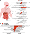
ENS Lewy pathology detected in postmortem studies of confirmed PD cases. (A,B) Each row represents one study, with the citation indicated. PD cases are to the right of the y-axis, while control cases are to the left of the y-axis. For each patient group, the number of cases with Lewy pathology (red) out of the total number of cases examined in that group is given next to the corresponding bar, with % positive for Lewy pathology given in parentheses. Only cases unique to each study are included. In (A), graphs (from top to bottom) represent studies of the esophagus, stomach, small intestine, large intestine, and rectum. In (B), for each study, the total number of PD or control cases with Lewy pathology detected in any gastrointestinal segment is shown. * Indicates an unknown number of subjects from these studies are overlapping. N.I., Not included in the study. N.S., Not shown in the study. Figure created using Biorender.com and GraphPad Prism 9. Refs. [9,11,12,13,14,15,16,17,18,19,20,22,23,56].
Figure 3
Figure 3
Gut-to-brain α-synuclein transmission hypothesis of PD. Evidence from human studies suggests that Lewy pathology in the ENS of PD patients is concentrated in the upper gastrointestinal tract, with the greatest LB density found in the esophagus and stomach. This distribution parallels vagal innervation of the ENS, consistent with a potential transmission of α-synuclein from the ENS retrogradely through the vagal nerve into the CNS. Once in the CNS, Lewy pathology appears to spread from the brainstem to the midbrain and finally to cortical regions, resulting in neurodegeneration and functional decline. Several unresolved questions relating to the gut-to-brain hypothesis of PD are highlighted. Figure created using Biorender.com.

Similar articles

Cited by

References

    1. Ding C., Wu Y., Chen X., Chen Y., Wu Z., Lin Z., Kang D., Fang W., Chen F. Global, regional, and national burden and attributable risk factors of neurological disorders: The Global Burden of Disease study 1990–2019. Front. Public Health. 2022;10:952161. doi: 10.3389/fpubh.2022.952161. - DOI - PMC - PubMed
    1. Forno L.S. Neuropathology of Parkinson’s disease. J. Neuropathol. Exp. Neurol. 1996;55:259–272. doi: 10.1097/00005072-199603000-00001. - DOI - PubMed
    1. Spillantini M.G., Schmidt M.L., Lee V.M., Trojanowski J.Q., Jakes R., Goedert M. Alpha-synuclein in Lewy bodies. Nature. 1997;388:839–840. doi: 10.1038/42166. - DOI - PubMed
    1. Hoehn M.M., Yahr M.D. Parkinsonism: Onset, progression and mortality. Neurology. 1967;17:427–442. doi: 10.1212/WNL.17.5.427. - DOI - PubMed
    1. Lim S.Y., Fox S.H., Lang A.E. Overview of the extranigral aspects of Parkinson disease. Arch. Neurol. 2009;66:167–172. doi: 10.1001/archneurol.2008.561. - DOI - PubMed