Review
. 2023 Apr 13;24(8):7208.
doi: 10.3390/ijms24087208.

Extracellular Vesicles in Breast Cancer: From Biology and Function to Clinical Diagnosis and Therapeutic Management

Affiliations
Review

Extracellular Vesicles in Breast Cancer: From Biology and Function to Clinical Diagnosis and Therapeutic Management

Sylvain Loric et al. Int J Mol Sci. .

Abstract

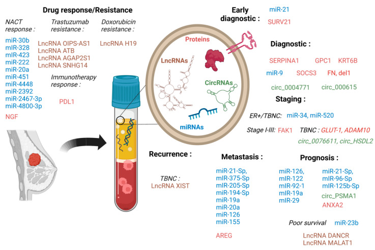
Breast cancer (BC) is the first worldwide most frequent cancer in both sexes and the most commonly diagnosed in females. Although BC mortality has been thoroughly declining over the past decades, there are still considerable differences between women diagnosed with early BC and when metastatic BC is diagnosed. BC treatment choice is widely dependent on precise histological and molecular characterization. However, recurrence or distant metastasis still occurs even with the most recent efficient therapies. Thus, a better understanding of the different factors underlying tumor escape is mainly mandatory. Among the leading candidates is the continuous interplay between tumor cells and their microenvironment, where extracellular vesicles play a significant role. Among extracellular vesicles, smaller ones, also called exosomes, can carry biomolecules, such as lipids, proteins, and nucleic acids, and generate signal transmission through an intercellular transfer of their content. This mechanism allows tumor cells to recruit and modify the adjacent and systemic microenvironment to support further invasion and dissemination. By reciprocity, stromal cells can also use exosomes to profoundly modify tumor cell behavior. This review intends to cover the most recent literature on the role of extracellular vesicle production in normal and cancerous breast tissues. Specific attention is paid to the use of extracellular vesicles for early BC diagnosis, follow-up, and prognosis because exosomes are actually under the spotlight of researchers as a high-potential source of liquid biopsies. Extracellular vesicles in BC treatment as new targets for therapy or efficient nanovectors to drive drug delivery are also summarized.

Keywords: breast cancer; diagnostic; exosomes; extracellular vesicles; prognosis; targeting; therapy; vector.


Conflict of interest statement

The authors declare no conflict of interest.

Figures

Figure 1
Figure 1
SEVs biogenesis and release. SEVs may have multiple origins. They can originate from plasma membrane budding, which leads to heterogeneous membranous medium-large vesicles (MLEVs) shedding. Small EVs (SEVs, exosomes) originate from the internal budding of plasma membranes giving rise to early endosomes. By complex maturating interactions with the Golgi apparatus (cargo-in/cargo-out), early endosomes become late ones. The membranes of late endosomes form intraluminal vesicles (ILVs), small cargos containing proteins from the plasma membrane, and Golgi as well as nucleic acids. Endosomal cargo sorting was performed through either ESCRT-dependent or -independent routes; the ESCRT complex being the key machinery of protein sorting into SEVs. ILVs are contained in multivesicular bodies (MVBs) that fuse with either plasma membrane (after Rab-driven docking), releasing SEVs in the extracellular space (through Snare complex assembly) or with lysosomes for further internal degradation.
Figure 2
Figure 2
Exosome membrane molecules and their cargo content. Small extracellular vesicles (SEVs) are nano-sized membrane vesicles released by a variety of cell types and are thought to play important roles in intercellular communications. SEVs contain many kinds of proteins, either cytosolic or plasma membrane ones. Transporters, receptors, and signaling proteins, but also enzymes, can be evidenced. Metabolites are also present as well as nucleic acids. Genomic and mitochondrial DNAs and multiple RNAs (mRNAs, miRNA, lncRNA, circRNA, etc.) can be detected. Through the horizontal transfer of these bioactive molecules, SEVS are emerging as local and systemic cell-to-cell mediators of oncogenic information.
Figure 3
Figure 3
Bidirectional communication between tumor cells and their surrounding environment. The tumor microenvironment (TME) is a complex and dynamic network that includes normal breast (NBC), tumor (TC), cancer-associated fibroblasts (CAFs), mesenchymal stromal (MSC), immune (tumor-associated macrophages TAM), and endothelial cells (EC). TC can bidirectionally signal to each other through SEVs production. TC can produce SEVs that will regulate MSCs’, CAFs’, and TAMs’ differentiation and activity. MSCs as well as TCs can regulate ECs’ activity, especially in hypoxic situations. TAMs, CAFs, and ECs can cooperate to promote angiogenesis. An antitumor immune response is largely modulated by BC cells through either extracellular signaling molecules (cytokines, etc.) secretion or SEVs production and release. BC cells SEVs contain inhibiting or activating molecules that favor target cells expansion, mobilization, and recruitment (CD4+ T cells (LT4), Tregs, and MSCs), polarization and activation (tumor-associated macrophages TAMs M2), and block others (CD8+ T cells (LT8), dendritic cells (DC), and natural killer NK cells).
Figure 4
Figure 4
SEVs cargo as relevant breast cancer biomarkers. Among all the molecules present in SEVs, only a subset (proteins, miRNAs, and LncRNAs) have been shown to be of potential clinical value in CRC detection, diagnosis, prognosis, and treatment response evaluation. All referenced markers were found to be differentially expressed in cancer patients and in healthy people: miRs are depicted in blue, LncRNAs in brown, CirRNAs in green, and proteins in pink. NACT: neoadjuvant chemotherapy.This figure was created with BioRender (DT2576K3SR agreement number).

References

    1. Sung H., Ferlay J., Siegel R.L., Laversanne M., Soerjomataram I., Jemal A., Bray F. Global Cancer Statistics 2020: GLOBOCAN Estimates of Incidence and Mortality Worldwide for 36 Cancers in 185 Countries. CA Cancer J. Clin. 2021;71:209–249. doi: 10.3322/caac.21660. - DOI - PubMed
    1. Ferlay J., Colombet M., Soerjomataram I., Dyba T., Randi G., Bettio M., Gavin A., Visser O., Bray F. Cancer Incidence and Mortality Patterns in Europe: Estimates for 40 Countries and 25 Major Cancers in 2018. Eur. J. Cancer. 2018;103:356–387. doi: 10.1016/j.ejca.2018.07.005. - DOI - PubMed
    1. Gennari A., André F., Barrios C.H., Cortés J., de Azambuja E., DeMichele A., Dent R., Fenlon D., Gligorov J., Hurvitz S.A., et al. ESMO Clinical Practice Guideline for the Diagnosis, Staging and Treatment of Patients with Metastatic Breast Cancer. Ann. Oncol. 2021;32:1475–1495. doi: 10.1016/j.annonc.2021.09.019. - DOI - PubMed
    1. Harbeck N., Penault-Llorca F., Cortes J., Gnant M., Houssami N., Poortmans P., Ruddy K., Tsang J., Cardoso F. Breast Cancer. Nat. Rev. Dis. Primers. 2019;5:66. doi: 10.1038/s41572-019-0111-2. - DOI - PubMed
    1. Luque-Bolivar A., Pérez-Mora E., Villegas V.E., Rondón-Lagos M. Resistance and Overcoming Resistance in Breast Cancer. Breast Cancer Targets Ther. 2020;12:211–229. doi: 10.2147/BCTT.S270799. - DOI - PMC - PubMed