. 2023 Apr 13;24(8):7209.
doi: 10.3390/ijms24087209.

Ursodeoxycholic Acid Binds PERK and Ameliorates Neurite Atrophy in a Cellular Model of GM2 Gangliosidosis

Affiliations

Ursodeoxycholic Acid Binds PERK and Ameliorates Neurite Atrophy in a Cellular Model of GM2 Gangliosidosis

Carolina Morales et al. Int J Mol Sci. .

Abstract

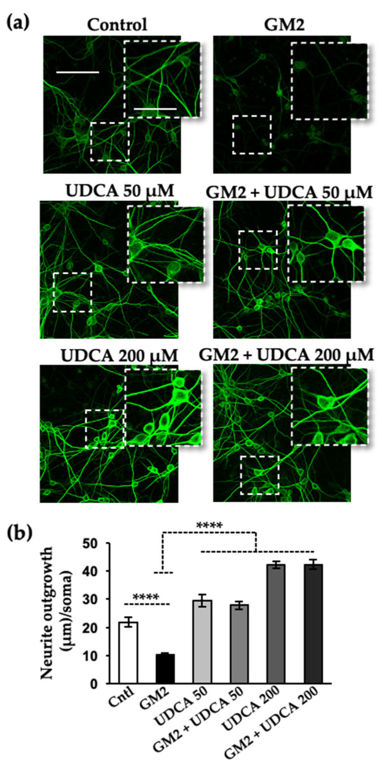
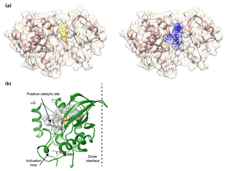
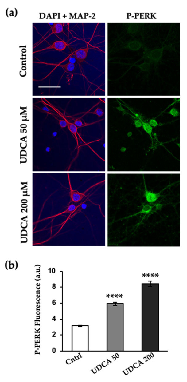
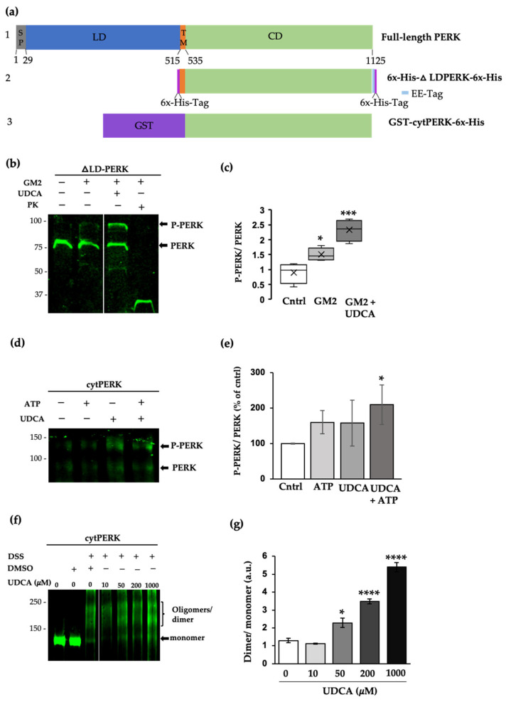
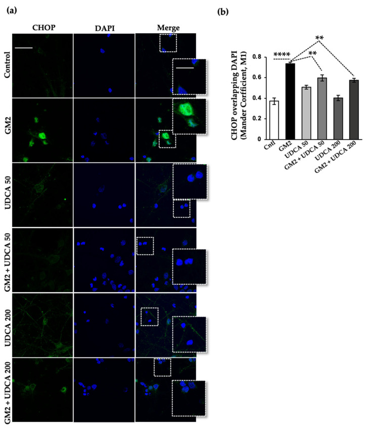
The Unfolded protein response (UPR), triggered by stress in the endoplasmic reticulum (ER), is a key driver of neurodegenerative diseases. GM2 gangliosidosis, which includes Tay-Sachs and Sandhoff disease, is caused by an accumulation of GM2, mainly in the brain, that leads to progressive neurodegeneration. Previously, we demonstrated in a cellular model of GM2 gangliosidosis that PERK, a UPR sensor, contributes to neuronal death. There is currently no approved treatment for these disorders. Chemical chaperones, such as ursodeoxycholic acid (UDCA), have been found to alleviate ER stress in cell and animal models. UDCA's ability to move across the blood-brain barrier makes it interesting as a therapeutic tool. Here, we found that UDCA significantly diminished the neurite atrophy induced by GM2 accumulation in primary neuron cultures. It also decreased the up-regulation of pro-apoptotic CHOP, a downstream PERK-signaling component. To explore its potential mechanisms of action, in vitro kinase assays and crosslinking experiments were performed with different variants of recombinant protein PERK, either in solution or in reconstituted liposomes. The results suggest a direct interaction between UDCA and the cytosolic domain of PERK, which promotes kinase phosphorylation and dimerization.

Keywords: ATP binding pocket; chemical chaperones; lysosomal storage disease.


Conflict of interest statement

The authors declare no conflict of interest. The funders had no role in the design of the study; in the collection, analyses, or interpretation of data; in the writing of the manuscript; or in the decision to publish the result.

Figures

Figure 1
Figure 1
UDCA treatment reverses neuritic atrophy induced by GM2 accumulation. (a) Primary cortical neurons (10 DIV) were loaded with 2 μM of GM2 or left unloaded; after 1 h of incubation, the cells were treated with either vehicle, 50 μM or 200 μM of UDCA for an additional 19 h. At the end of this incubation, the cells were fixed with 4% paraformaldehyde, immunolabeled with mouse anti-MAP-2 antibody, and visualized with Alexa Fluor-488 conjugated secondary antibody. Images were recorded with a confocal microscope (Zeiss LSM 800). Scale bars: 100 μm (regular images), 50 μm (magnified images); (b) Histogram (mean ± SEM) represents neurite outgrowth with respect to total cells, analyzed with Image J plug-ins (NIH, USA). **** p ≤ 0.00001 with respect to the control as determined by one-way ANOVA, n = 3.
Figure 2
Figure 2
Docking models obtained on HADDOCK. (a) Left, model of mPERK bound to AMP-PNP, obtained on Chimera by combining the structures of PERK (3QD2) and PRK-AMP-PNP (2A19). For AMP-PNP, the surface appears yellow and sticks are colored by atom. Right, superposition of the surfaces of the four best docking models is shown in blue. Sticks colored by atom correspond to UDCA molecules; (b) Overall structure of the mPERK monomer. The threonine (Thr) 980 and the activation loop are shown. The α–G helix, which provides the docking site for eIF2α, is also depicted. Note that the binding site for UDCA matches the putative catalytic site.
Figure 3
Figure 3
PERK phosphorylation levels are increased by UDCA treatment. (a) Primary cortical primary neurons (10 DIV) were incubated with or without 50 μM and 200 μM of UDCA for 19 h. The cells were fixed, immunolabeled with an anti-P-PERK antibody, and visualized with Alexa Fluor-488 conjugated secondary antibody. Nuclei were stained with DAPI (blue) and microtubule-associated protein 2 with chicken anti-MAP-2 antibody and visualized with Alexa-Fluor-594. Images were recorded with a confocal microscope. Scale bars: 30 μm; (b) Histogram (mean ± SEM) represents phospho-PERK fluorescence intensity, analyzed with Image J plug-ins (NIH, USA). **** p ≤ 0.00001 with respect to the control as determined by one-way ANOVA, n = 3.
Figure 4
Figure 4
UDCA directly interacts with PERK and prompts its phosphorylation and oligomerization. (a) Diagrams: (1) Full-length mouse PERK provided as a reference; residue numbering based on UniProt Q9Z2B5. The signal peptide (SP) and the luminal (LD), transmembrane (TM), and cytosolic (CD) domains are indicated. (2) Hexahistidine-tagged PERK protein, lacking the luminal domain (ΔLD-PERK), was expressed in E. coli and used to constitute proteoliposomes. (3) The cytosolic domain of PERK (cytPERK) expressed in the bacteria as a GST fusion protein, and used in bulk solution reactions; (b) Immunodetection of PERK using an antibody that recognizes both the phosphorylated and non-phosphorylated forms. His-tagged ΔLD-PERK was incorporated into control or GM2 liposomes, purified by ultracentrifugation, and incubated with ATP (1 µM), in the presence or absence of UDCA (200 µM) for 20 min at 27 °C. Proteins were resolved on 10% SDS-PAGE; (c) Boxplots indicate median, 25th and 75th percentile limits, and extreme PERK phosphorylation values determined by the phosphorylated to non-phosphorylated ratio; (d) GST-cytPERK was incubated with ATP (0.1 mM), resolved on 7% SDS-PAGE, and probed with an anti-PERK antibody as in (b); (e) Histogram corresponding to densitometric analysis of P-PERK and PERK values normalized with control ratio value (100%); (f) Recombinant GST-cytPERK (1.3 µM) was incubated in the presence of DSS (0.4 mM) or DMSO (vehicle) and increasing concentrations of UDCA for 30 min at 27 °C. The reaction was quenched with 50 mM Tris-HCl (15 min at 27 °C). The proteins were resolved on 7% SDS-PAGE and probed as in (b); (g) Histogram corresponding to densitometric analysis of ratio values for dimer relative to monomer (c) * p ≤ 0.05, *** p ≤ 0.0001, n = 3; (e) * p ≤ 0.05, n = 3; (g) * p ≤ 0.05, **** p ≤ 0.00001, n = 4 (ANOVA, Tukey’s HSD test).
Figure 5
Figure 5
UDCA decreases the translocation of the transcription factor CHOP to the nuclei in GM2-stressed neurons. (a) Primary cortical neurons (10 DIV) were loaded with GM2, then treated with UDCA as in Figure 1. Fixed cells were labeled with anti-CHOP antibody (green) and their nuclei were labeled with DAPI (blue). Images were recorded with a confocal microscope. Scale bars: 30 μm (regular images), 15 μm (magnified images); (b) Histogram (mean ± SEM) represents Manders’ overlap coefficient, M1 (Channel 1: green, Channel 2: blue) calculated on Image J (NIH, USA). ** p ≤ 0.001; **** p ≤ 0.00001 with respect to the control as determined by one-way ANOVA, n = 3.

Similar articles

Cited by

References

    1. Van Ziel A.M., Scheper W. The UPR in Neurodegenerative Disease: Not Just an Inside Job. Biomolecules. 2020;10:1090. doi: 10.3390/biom10081090. - DOI - PMC - PubMed
    1. Moreno J.A., Radford H., Peretti D., Steinert J.R., Verity N., Martin M.G., Halliday M., Morgan J., Dinsdale D., Ortori C.A., et al. Sustained translational repression by eIF2alpha-P mediates prion neurodegeneration. Nature. 2012;485:507–511. doi: 10.1038/nature11058. - DOI - PMC - PubMed
    1. Hetz C., Saxena S. ER stress and the unfolded protein response in neurodegeneration. Nat. Rev. Neurol. 2017;13:477–491. doi: 10.1038/nrneurol.2017.99. - DOI - PubMed
    1. Lin J.H., Li H., Yasumura D., Cohen H.R., Zhang C., Panning B., Shokat K.M., Lavail M.M., Walter P. IRE1 signaling affects cell fate during the unfolded protein response. Science. 2007;318:944–949. doi: 10.1126/science.1146361. - DOI - PMC - PubMed
    1. Lin J.H., Li H., Zhang Y., Ron D., Walter P. Divergent effects of PERK and IRE1 signaling on cell viability. PLoS ONE. 2009;4:e4170. doi: 10.1371/journal.pone.0004170. - DOI - PMC - PubMed