. 2023 Apr 13;24(8):7226.
doi: 10.3390/ijms24087226.

Investigation of the Compatibility between Warheads and Peptidomimetic Sequences of Protease Inhibitors-A Comprehensive Reactivity and Selectivity Study

Affiliations

Investigation of the Compatibility between Warheads and Peptidomimetic Sequences of Protease Inhibitors-A Comprehensive Reactivity and Selectivity Study

Patrick Müller et al. Int J Mol Sci. .

Abstract

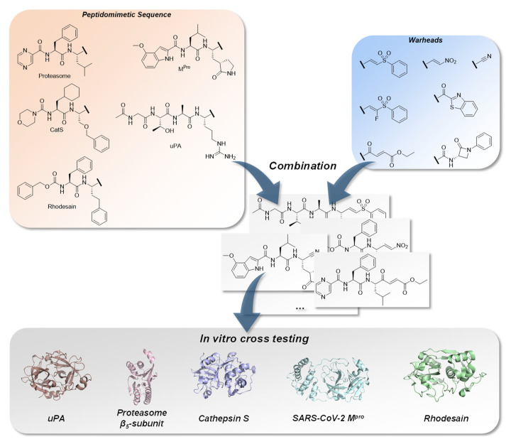
Covalent peptidomimetic protease inhibitors have gained a lot of attention in drug development in recent years. They are designed to covalently bind the catalytically active amino acids through electrophilic groups called warheads. Covalent inhibition has an advantage in terms of pharmacodynamic properties but can also bear toxicity risks due to non-selective off-target protein binding. Therefore, the right combination of a reactive warhead with a well-suited peptidomimetic sequence is of great importance. Herein, the selectivities of well-known warheads combined with peptidomimetic sequences suited for five different proteases were investigated, highlighting the impact of both structure parts (warhead and peptidomimetic sequence) for affinity and selectivity. Molecular docking gave insights into the predicted binding modes of the inhibitors inside the binding pockets of the different enzymes. Moreover, the warheads were investigated by NMR and LC-MS reactivity assays against serine/threonine and cysteine nucleophile models, as well as by quantum mechanics simulations.

Keywords: covalent inhibitors; in vitro study; peptidomimetic sequence; protease inhibitors; reactivity and selectivity study; warhead.


Conflict of interest statement

The authors declare no conflict of interest.

Figures

Figure 1
Figure 1
Combination of characteristic peptidomimetic inhibitor sequences for the targets: urokinase-type plasminogen activator (uPA), PDB-ID: 1W10 [41], proteasome β5-subunit, PDB-ID: 5LF3 [42], cathepsin S, PDB-ID: 1MS6 [43], SARS-CoV-2 main protease (Mpro), PDB-ID: 6XR3 [44] and rhodesain, PDB-ID: 2P7U [45], with selected warheads (vinyl sulfone, F-vinyl sulfone, nitroalkene, α-ketobenzothiazole, 4-oxoenoate, nitrile and β-lactam). The resulting compounds were tested on each target to determine affinity and selectivity.
Scheme 1
Scheme 1
Synthesis of precursor molecules. (A) Synthesis of phosphonate building block 4. (B) Synthesis of β-Lactam building block 9. Reaction conditions: (a) n-BuLi, DECP, THF, −78 °C; (b) 3, LHMDS, Selectfluor®, THF, DMF, −78 °C, 3 h, 49%; (c) Cbz-Cl, NaHCO3, H2O, 12 h, rt, 90%; (d) aniline, TBTU, HOBt · H2O, EtOAc, 12 h, rt, 74%; (e) ImSO2, NaH, DMF, F20 °C, 1.5 h, 77%; (f) Pd/C, H2, THF, 88%.
Scheme 2
Scheme 2
Synthesis of rhodesain compounds. Reaction conditions: (a) N,O-dimethylhydroxylamine · HCl, DCC, HOBt · H2O, DIPEA, THF, rt, 12 h, 46%; (b) LAH, THF, 0 °C, 2 h, 67%; (c) 1. MeNO2, Et3N, DCM, rt, 8 h, 2. TFA, DCM, rt, 0.5 h, 3. Cbz-(l)Phe-OH, EDC · HCl, TEA, DCM, rt, 12 h, 4. MsCl, DIPEA, DCM, rt, 2 h, 75%; (d) 3/4, LiHMDS, THF, −80 °C, 12 h, 59% (14), 57% (15); (e) TFA, DCM, Cbz-(l)Phe-OH, T3P, DIPEA, DMF, rt, 12 h, 65% (16), 48% (17); (f) 1. benzothiazole, n-BuLi, THF, −78 °C, 3 h, 2. TFA, DCM, rt, 2 h, 3. Cbz-(l)Phe-OH, EDC · HCl, HOBt · H2O, Et3N, DCM, rt, 8 h, 56%; (g) H-(l)hPhe-OMe, HATU, 2,4,6-collidine, DCM/DMF, rt, 16 h, quant.; (h) DMMP, n-BuLi, THF, –70 °C, 2 h, 98%; (i) ethyl glyoxylate, K2CO3, EtOH, rt, 2 h, 76%; (j) LiOH, THF/H2O, rt, 16 h, quant.; (k) 9, HATU, 2,4,6-collidine, DCM/DMF, rt, 16 h, 67%; (l) 1. EDC · HCl, HOBt · H2O, NH4OH, DMF, rt, 12 h, 2. TFAA, pyridine, DMF, 0 °C, 0.1 h, 40%.
Scheme 3
Scheme 3
Synthesis of cathepsin S compounds. Reaction conditions: (a) SOCl2, MeOH, –10 °C, 16 h, 91%; (b) morpholine, triphosgene, NaHCO3, CHCl2, 0 °C; 16 h, 98%; (c) LiOH, THF/H2O, 3 h, 97%; (d) 1. NaCN, NH4Cl, NH3, 2-(benzyloxy)acetaldehyde, Et2O, 2. HATU, 2,4,6-collidine, DCM/DMF, rt, 16 h, 53%; (e) N,O-dimethylhydroxylamine · HCl, DCC, HOBt · H2O, DIPEA, THF, –15–0°C, 16 h, 80%; (f) 1. LAH, Et2O, 0 °C, 2 h, 2. 3/4, KHMDS/ LHMDS, THF, –78 °C, 3 h; 75% (33), 44% (34); (g) 1. 4 N HCl in 1,4-dioxane, 2. 29, HATU, collidine, DCM/DMF, rt, 16 h, 63% (35), 60% (36); (h) MeI, DMF, 0 °C, 16 h, 97%; (i) n-BuLi, DMMP, THF, –78 °C, 3 h, 79%; (j) 1. 4 N HCl in 1,4-dioxane, 2. 29, HATU, 2,4,6-collidine, DCM/DMF, rt, 16 h, 52%; (k) LiCl, ethyl glyoxylate, DIPEA, MeCN, 0 °C, 2 h, 39%; (l) benzothiazole, n-BuLi, THF, –78 °C, 3 h, 38%; (m) 1. 4 N HCl in 1,4-dioxane, 2. 29 HATU, 2,4,6-collidine, DCM/DMF, rt, 16 h, 43%; (n) 1.4 N HCl in 1,4-dioxane, 2. 29, HATU, collidine, DCM/DMF, rt, 16 h, 90%; (o) LiOH, THF/H2O, rt, 3 h, 99%; (p) 9, HATU, 2,4,6-collidine, DCM/DMF, rt, 16 h, 81%; (q) NaBH4, MeOH, THF, 0 °C, 16 h, 91%; (r) Dess–Martin–Periodinan, DCM, rt, 16 h, 70%; (s) NaH, MeNO2, THF, 0 °C, 1 h, 58%; (t) MsCl, Et3N, DCM, 0 °C, 3 h, 45%.
Scheme 4
Scheme 4
Synthesis of proteasome β5-subunit compounds. Reaction conditions: (a) N,O-dimethylhydroxylamine · HCl, TBTU, HOBt · H2O, 2,4,6-collidine, DCM, 0–20 °C, 16 h, 86%; (b) LAH, THF, 0 °C, 30 min, 29%; (c) 4, LiCl, DBU, MeCN, 0 °C, 1 h, 80%; (d) 1. 4 N HCl in 1,4-dioxane, rt, 1 h, 2. Boc-Phe-OH, TBTU, HOBt · H2O, 2,4,6-collidine, DCM, 0 °C, 16 h, 97%; (e) 1. 4 N HCl in 1,4-dioxane, rt, 1.5 h, 2. pyrazinoic acid, TBTU, HOBt · H2O, 2,4,6-collidine, DCM, 0 °C, 16 h, 67%; (f) 1. 4 N HCl in 1,4-dioxane, rt, 1 h, 2. Boc-(l)Phe-OH, TBTU, HOBt · H2O, 2,4,6-collidine, DCM, 0–20 °C, 16 h, 97%; (g) 1. 4 N HCl in 1,4-dioxane, rt, 1 h, 2. pyrazinoic acid, TBTU, HOBt · H2O, 2,4,6-collidine, DCM, 0–20 °C, 16 h, 88%; (h) 1. LAH, THF, 0 °C, 1 h, 2. 3, LiCl, DBU, MeCN, 0 °C, 1.5 h, 11%; (i) 1. LAH, THF, 0 °C, 1 h, 2. MeNO2 Et3N, DCM, 0–20 °C, 16h, 3. MsCl, DIPEA, DCM, rt, 3 h, 19%; (j) benzothiazole, n-BuLi, THF, –78 °C, 6 h, 65%; (k) 1. 4 N HCl in 1,4-dioxane, rt, 1 h, 2. Boc-(l)Phe-OH, TBTU, HOBt · H2O, 2,4,6-collidine, DCM, 0–20 °C, 16 h, 68%; (l) 1. 4 N HCl in 1,4-dioxane, rt, 1 h, 2. pyrazinoic acid, TBTU, HOBt · H2O, 2,4,6-collidine, DCM, 0–20 °C, 16 h, 53%; (m) Boc2O, NaHCO3, water, 1,4-dioxane, 3 h, rt, 99%; (n) n-BuLi, THF, –78 °C, 6 h, 88%; (o) 1. 4 N HCl in 1,4-dioxane, rt, 1 h, 2. Boc-(l)Phe-OH, TBTU, HOBt · H2O, 2,4,6-collidine, DCM, DMF, 0–20 °C, 16 h, 72%; (p) 1. 4 N HCl in 1,4-dioxane, rt, 1 h, 2. pyrazinoic acid, TBTU, HOBt · H2O, 2,4,6-collidine, DCM, DMF, 0–20 °C, 16 h, 66%; (q) ethyl glyoxylate, LiCl, DIPEA, MeCN, 1 h, 0 °C, 79%; (r) Boc-(l)Phe-OH, TBTU, HOBt · H2O, 2,4,6-collidine, DCM, 0–20 °C, 16 h, 82%; (s) 1. 4 N HCl in 1,4-dioxane, rt, 1 h, 2. pyrazinoic acid, TBTU, HOBt · H2O, 2,4,6-collidine, DCM, 0–20 °C, 16 h, 82%; (t) LiOH, water, THF, rt, 17 h, quant.; (u) 9, TBTU, HOBt · H2O, 2,4,6-collidine, DCM, 0–20 °C, 16 h, 65%; (v) 7 N NH3 in MeOH, rt, 48 h, 89%; (w) cyanuric chloride, DMF, 0 °C, 48 h, 47%.
Scheme 5
Scheme 5
Synthesis of SARS-CoV-2 Mpro compounds. Reaction conditions: (a) H-(l)Leu-OBn · pTsOH, TBTU, DIPEA, DCM, 0–20 °C, 16 h, 93%; (b) H2, Pd/C (10%), EtOH, quant.; (c) DMMP, n-BuLi, THF, –78 °C, 5 h, 24%; (d) 1. TFA, DCM, 0 °C, 3 h, 2. 77, EtOCOCl, NMM, THF, –20 °C, 2 h, 40%; (e) ethyl glyoxylate, LiCl, DIPEA, MeCN, 0 °C, 2 h, 47%; (f) 1. TFA, DCM, 0 °C, 3 h, 2. 77, HATU, 2,4,6-collidine, DMF, 0–20 °C, 16 h, 83%; (g) 1. LiOH, THF/H2O, 0–4 °C, 16 h, 2. NH4OH, HATU, OxymaPure®, 2,4,6-colllidine, DMF, 0–20 °C, 16 h, 65%; (h) burgess reagent, DCM, rt, 2 h, 67%; (i) 1. LiOH, THF/MeOH/H2O, 0–4 °C, 16 h, 2. N,O-dimethylhydroxylamine · HCl, HATU, 2,4,6-colllidine, DMF, 0–20 °C, 16 h, 76%; (j) 1. LAH, THF, −20 °C, 2 h, 2. MeNO2, EtN3, DCM, rt, 15 h, 71%; (k) 1. TFA, DCM, 0 °C, 3 h, 2. 77, EDC · HCl, HOBt · H2O, DIPEA, DCM, 0–20 °C, 16 h, 3. MsCl, DIPEA, DCM, 0–20 °C, 16 h, 50%; (l) benzothiazole, n-BuLi, THF, –78 °C, 5 h, 59%; (m) 1. TFA, DCM, 0 °C, 3 h, 2. 77, EtOCOCl, NMM, THF, –20 °C, 2 h, 54%; (n) 1. LAH, THF, −20 °C, 2 h, 2. 3/4, LiCl, DBU, MeCN, 0 °C, 2 h, 65% (89), 35% (90); (o) 1. TFA, DCM, 0 °C, 3 h, 2. 77, EtOCOCl, NMM, THF, –20 °C, 2 h, 18% (91), 15% (92); (p) 1. LiOH, water, THF, 16 h, 2. 9, TBTU, HOBt · H2O, DIPEA, DCM, 0–20 °C, 48 h, 39%; (q) TFA, DCM, 0 °C, 3 h, 2. 77, EtOCOCl, NMM, THF, –20 °C, 2 h, 80%.
Scheme 6
Scheme 6
Synthesis of the uPA compounds. (A) solid phase peptide synthesis of building block 99. (B) combined synthesis of the final uPA compounds. Reaction conditions: (a) N,O-dimethylhydroxylamine· HCl, TBTU, DIPEA, DCM, rt, 12 h, 95%; (b) benzothiazole, n-BuLi, THF, –78 °C, 2 h, 76%; (c) 1. TFA, DCM, rt, 0.5 h, 2. HATU, DIPEA, DMF, DCM, rt, 12 h, 3. TFA, DCM, rt, 2 h, 10%; (d) 1. LAH, THF, 0 °C, 2. 3/4, LiCl, DBU, MeCN, 0 °C, 1 h, 72% (104), 31% (105); (e) 1. TFA, DCM, rt, 0.5 h, 2. 99, HATU, DIPEA, DMF, DCM, rt, 12 h, 3. TFA, DCM, rt, 2 h, 16% (106), 11% (107).
Scheme 7
Scheme 7
Synthesis of the reactivity probes. Reaction conditions: (a) 9, HATU, collidine, DCM, DMF, rt, 16 h, 97%; (b) TBTU, HOBt · H2O, N,O-dimethylhydroxylamine · HCl, 2,4,6-collidine, rt, 12 h, quant.; (c) 1. LAH, Et2O, 0 °C, 1 h, 89%; (d) 3/4, LiCl, DBU, MeCN, 0 °C, 1 h, 50% (109), 30% (110); (e) 1. NaH, MeNO2, THF, 0 °C, 1 h, 2. MsCl, Et3N, DCM, 0 °C, 1 h, 15%; (f) DMMP, n-BuLi, THF, –78 °C, 1.5 h, 70%; (g) ethyl glyoxylate, LiCl, DBU, MeCN, 0 °C, 1 h, 45%; (h) TBTU, HOBt · H2O, N,O-dimethylhydroxylamine · HCl, 2,4,6-collidine, 0–20 °C, 16 h, 95%; (i) benzothiazole, n-BuLi, THF, –78 °C, 3.5 h, 56%; (j) EDC · HCl, HOBt · H2O, NH4OH, DMF, rt, 16 h, 14%; (k) TFAA, pyridine, THF, –10 °C, 2 h, 74%.
Scheme 8
Scheme 8
Reaction scheme of the reactivity assay with both model nucleophiles under equal reaction conditions and the vinyl sulfone moiety 109.
Figure 2
Figure 2
1H-NMR spectra of the 4-oxoenoate 112 before the addition (0 min) and after 5, 30, 60, 120 and 240 min reaction time with 2-phenylethanthiol in the absence of triethylamine. The integrals of the β-proton of the double bond at 7.17 ppm in relation to the 2-CH2 signal of the internal standard 1,3-dioxolane at 5.3 ppm are given. LC-MS analysis of the same reaction at 30 min.
Figure 3
Figure 3
LC-MS spectra of the β-lactam 108 before (0 min) the addition and after 5, 30 and 60 min reaction time with sodium ethoxide.
Figure 4
Figure 4
Progress curves of the reactions of the different warhead compounds with the model nucleophiles PhEtSH, PhEtSH + Et3N and EtONa as measured by NMR and LC-MS analysis.
Figure 5
Figure 5
(A) Free energies of the reactions for all inhibitor warheads with MeSH, MeSH + Et3N and MeO + 3H2O, computed as described in the Supplementary Materials section and Figures S24 by ωB97X−D/6−311++G**//ωB97X−D/6-31+G* calculations. (B) Free energy reaction paths of the α-ketobenzothiazole warhead with MeSH (red) and with MeSH in the presence of three water molecules (blue). For MeSH (red), the van der Waals complex (P vdw) and separated product molecules (P) are identical since the reaction yields only a single product molecule. For MeSH + water (blue), the energies are referenced on MeSH + 2H2O and the α-ketobenzothiazole warhead + H2O (R). (C) Free energy reaction paths of the β-lactam warhead with MeSH + NEt3 (red) and with MeO in the presence of three water molecules (blue). The reaction proceeds in three consecutive steps: first, nucleophilic attack at the amide carbonyl group (TS1); second, the opening of the lactam ring (TS2); and third, the proton transfer from the base (NEt3 or H2O) to the former amide nitrogen (TS3).
Figure 6
Figure 6
Inhibition data for the assays with uPA (A), proteasome β5-subunit (B), cathepsine S (C), SARS-CoV-2 Mpro (D) and rhodesain (E). pKi values were calculated from the Ki values (–log10(Ki/M)) [59]. The height of a bar indicates the inhibitory potency of an inhibitor towards the target enzyme, and the color of a bar indicates the warhead of the inhibitor; the peptidomimetic sequence is indicated by the enzyme name under the bars, e.g., the purple bar with the height value of 7.5 for rhodesain inhibition (Figure 6E) by inhibitor 40 with the oxoenoate warhead and the CatS sequence. * Respective compounds were inactive in the in vitro assay due to instability towards DTT in the buffer.
Figure 6
Figure 6
Inhibition data for the assays with uPA (A), proteasome β5-subunit (B), cathepsine S (C), SARS-CoV-2 Mpro (D) and rhodesain (E). pKi values were calculated from the Ki values (–log10(Ki/M)) [59]. The height of a bar indicates the inhibitory potency of an inhibitor towards the target enzyme, and the color of a bar indicates the warhead of the inhibitor; the peptidomimetic sequence is indicated by the enzyme name under the bars, e.g., the purple bar with the height value of 7.5 for rhodesain inhibition (Figure 6E) by inhibitor 40 with the oxoenoate warhead and the CatS sequence. * Respective compounds were inactive in the in vitro assay due to instability towards DTT in the buffer.
Figure 7
Figure 7
Predicted binding modes and polar interactions (yellow dashed lines) of different inhibitor classes with different enzymes (white carbon atoms and surface). For non-covalent docking poses, the distance between electrophilic carbon and nucleophilic sulfur or oxygen is shown as a red dashed line, with the distance measured in Å. For a clear view, only amino acids that form polar interactions with the ligands are shown as sticks and labelled. Black dashed lines indicate subpocket locations. Non-covalent docking poses are shown with cyan C-atoms and the covalent docking poses with green C-atoms. (A) Superposition of the non-covalent and the covalent docking pose of 13 with rhodesain, PDB-ID (2P7U). (B) Superposition of the non-covalent and the covalent docking pose of 30 with CatS, PDB-ID (1MS6). (C) Superposition of the non-covalent and the covalent docking pose of 90 with SARS-CoV-2-Mpro, PDB-ID (6XR3). (D) Predicted binding mode of non-covalently docked 103 with uPA, PDB-ID (1W10). (E) Superposition of the non-covalent docking pose of 72 and bortezomib (palegreen C-atoms) with the β-5 subunit of human 20S-proteasome, PDB-ID (5LF3).

Similar articles

Cited by

References

    1. López-Otín C., Overall C.M. Protease degradomics: A new challenge for proteomics. Nat. Rev. Mol. Cell Biol. 2002;3:509–519. doi: 10.1038/nrm858. - DOI - PubMed
    1. Grozdanić M., Vidmar R., Vizovišek M., Fonović M. Degradomics in Biomarker Discovery. Proteom. Clin. Appl. 2019;13:1800138. doi: 10.1002/prca.201800138. - DOI - PubMed
    1. Ruggiano A., Ramadan K. DNA–protein crosslink proteases in genome stability. Commun. Biol. 2021;4:11. doi: 10.1038/s42003-020-01539-3. - DOI - PMC - PubMed
    1. Lee C.W., Stankowski J.N., Chew J., Cook C.N., Lam Y.W., Almeida S., Carlomagno Y., Lau K.F., Prudencio M., Gao F.B., et al. The lysosomal protein cathepsin L is a progranulin protease. Mol. Neurodegener. 2017;12:55. doi: 10.1186/s13024-017-0196-6. - DOI - PMC - PubMed
    1. Eatemadi A., Aiyelabegan H.T., Negahdari B., Mazlomi M.A., Daraee H., Daraee N., Eatemadi R., Sadroddiny E. Role of protease and protease inhibitors in cancer pathogenesis and treatment. Biomed. Pharmacother. 2017;86:221–231. doi: 10.1016/j.biopha.2016.12.021. - DOI - PubMed

MeSH terms

LinkOut - more resources