. 2023 Apr 14;24(8):7243.
doi: 10.3390/ijms24087243.

Developmental Stage-Dependent Changes in Mitochondrial Function in the Brain of Offspring Following Prenatal Maternal Immune Activation

Affiliations

Developmental Stage-Dependent Changes in Mitochondrial Function in the Brain of Offspring Following Prenatal Maternal Immune Activation

Magdalena Cieślik et al. Int J Mol Sci. .

Abstract

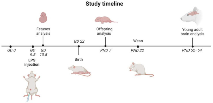
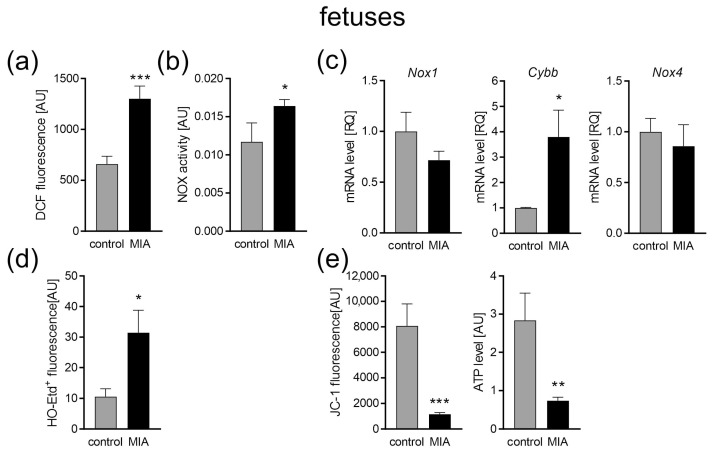
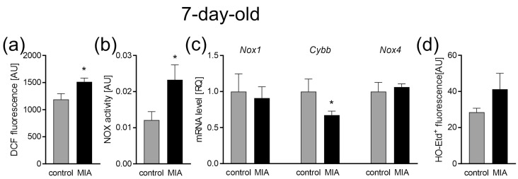
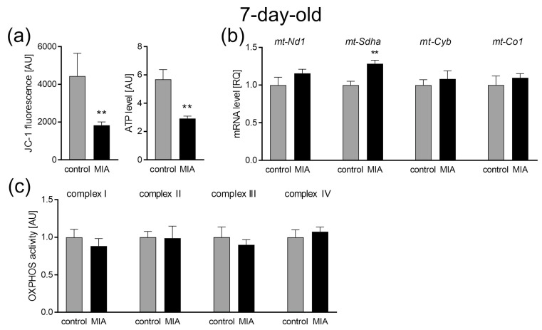
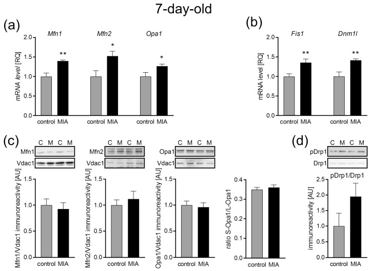
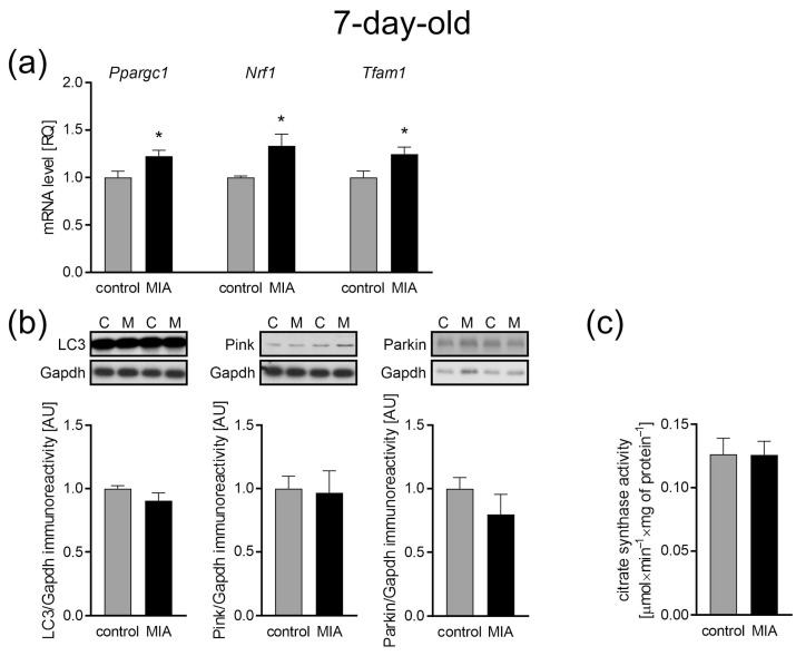
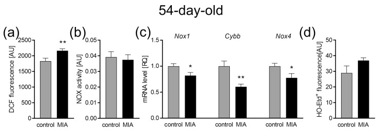
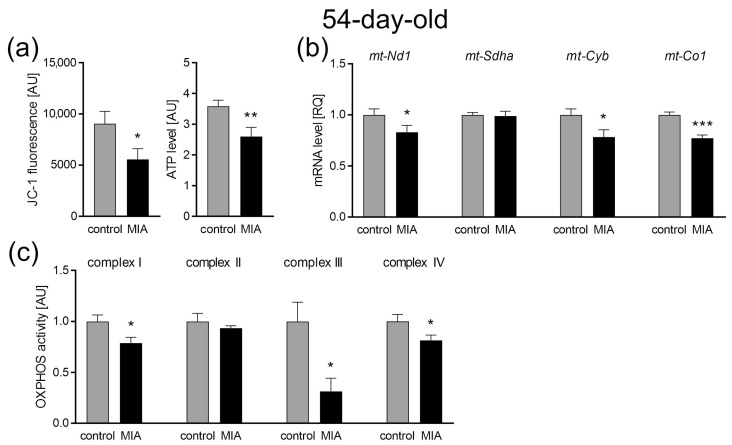
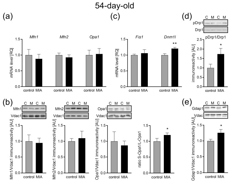
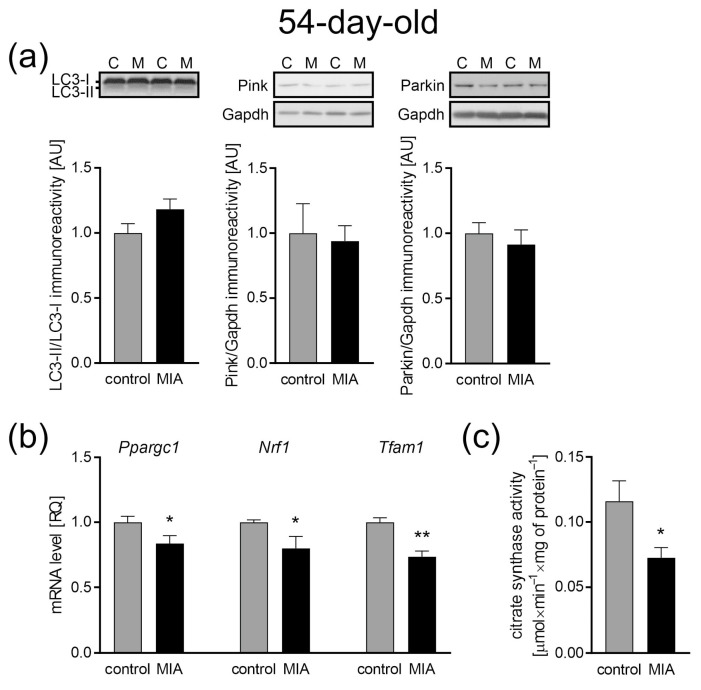
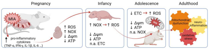
Maternal immune activation (MIA) is an important risk factor for neurodevelopmental disorders such as autism. The aim of the current study was to investigate the development-dependent changes in the mitochondrial function of MIA-exposed offspring, which may contribute to autism-like deficits. MIA was evoked by the single intraperitoneal administration of lipopolysaccharide to pregnant rats at gestation day 9.5, and several aspects of mitochondrial function in fetuses and in the brains of seven-day-old pups and adolescent offspring were analyzed along with oxidative stress parameters measurement. It was found that MIA significantly increased the activity of NADPH oxidase (NOX), an enzyme generating reactive oxygen species (ROS) in the fetuses and in the brain of seven-day-old pups, but not in the adolescent offspring. Although a lower mitochondrial membrane potential accompanied by a decreased ATP level was already observed in the fetuses and in the brain of seven-day-old pups, persistent alterations of ROS, mitochondrial membrane depolarization, and lower ATP generation with concomitant electron transport chain complexes downregulation were observed only in the adolescent offspring. We suggest that ROS observed in infancy are most likely of a NOX activity origin, whereas in adolescence, ROS are produced by damaged mitochondria. The accumulation of dysfunctional mitochondria leads to the intense release of free radicals that trigger oxidative stress and neuroinflammation, resulting in an interlinked vicious cascade.

Keywords: NADPH oxidase; ROS; animal model; autism; maternal immune activation; mitochondria; neurodevelopmental disorders.


Conflict of interest statement

The authors declare no conflict of interest.

Figures

Figure 1
Figure 1
Timeline of the study. LPS (100 µg/kg b.w.) was injected intraperitoneally (i.p.) into pregnant rats at gestation day 9.5 (GD 9.5), and the control animals received a single i.p. dose of solvent (sterile 0.9% NaCl). Some of the pregnant mothers were sacrificed 24 h after the LPS administration (GD 10.5). The fetuses were collected for further research. The other dams were allowed to give birth and nurture offspring under normal conditions. The day of birth was recorded as postnatal day (PND) 1. On PND 7, each litter was equalized to 10 (both male and female) and the eliminated pups were sacrificed, their brains were removed, and they were taken for further analysis. On PND 22 to 23, rat pups were separated and housed in groups of 3 or 4. Adolescent males were sacrificed at PND 52–54, their brains were removed, and the cerebral cortex was isolated for further analysis. The figure was created with BioRender.com.
Figure 2
Figure 2
The effect of MIA on oxidative stress and mitochondrial function in fetuses. Twenty-four hours after maternal LPS administration, rat fetuses were sacrificed and whole brains were collected. (a) The level of ROS was determined with a DCFH-DA probe (n = 8). (b) The activity of NADPH oxidase (NOX) was determined by the initial rate of inhibitable ferricytochrome c reduction (n = 5 and 7). (c) The mRNA levels of NOX subunits Nox1, Cybb, and Nox4 were determined using real-time PCR and were calculated by the ΔΔCt method with Actb (β-actin) as a reference gene (n = 6 and 5). (d) The generation of superoxide radicals was determined by the fluorimetric method using DHE (n = 4). (e) The mitochondrial membrane potential was determined by the fluorometric method using JC-1 and the mitochondrial ATP level was determined using the bioluminescence assay (n = 9 and 8). Data represent the mean value ± S.E.M. and were analysed using Student’s t-test, * p < 0.05, ** p < 0.01, *** p < 0.001, compared with the control group.
Figure 3
Figure 3
The effect of MIA on oxidative stress in the brain of 7-day-old offspring. (a) The level of reactive oxygen species was determined with a DCFH-DA probe (n = 8). (b) The level of NADPH oxidase (NOX) activity (n = 7 and 8). (c) The mRNA levels of NOX subunits Nox1, Cybb, and Nox4 were determined using a real-time PCR method with Actb (β-actin) as a reference gene (n = 5 and 6). (d) The generation of superoxide radicals was determined by the fluorimetric method using DHE (n = 4). Data represent the mean value ± S.E.M. and were analysed using Student’s t-test, * p < 0.05, compared with the control group.
Figure 4
Figure 4
The effect of MIA on mitochondrial function in the brain of 7-day-old MIA-offspring. (a) Mitochondrial membrane potential was determined by the fluorometric method using JC-1 and the mitochondrial ATP level was determined using the bioluminescence assay (n = 7 and 6). (b) The levels of mRNA for four mitochondrial electron transport chain complexes were analysed by real-time PCR using Actb as a reference gene: mt-Nd1 (n = 4 and 5), mt-Sdha (n = 4 and 5), mt-Cyb (n = 4 and 5), and mt-Co1 (n = 4 and 5). (c) The activity of respiratory complexes was measured using the kinetic spectrophotometric method: complex I (n = 7 and 5), complex II (n = 9), complex III (n = 5 and 4), and complex IV (n = 5). Data represent the mean value ± S.E.M. and were analysed using Student’s t-test. ** p < 0.01, compared with the control group.
Figure 5
Figure 5
The effect of MIA on the mitochondria fusion and fission in the brain of 7-day-old offspring rats. (a) The level of mRNA for Mfn1 (n = 4 and 5), Mfn2 (n = 5), and Opa1 (n = 5) was measured by real-time PCR with Actb as a reference gene. (b) The level of mRNA for Fis1 (n = 5) and Dnm1l (n = 5) was measured by real-time PCR with Actb as a reference gene. (c) The level of immunoreactivity of Mfn1 (n = 7 and 6), Mfn2 (n = 7 and 8), Opa1, and S-Opa1/L-Opa1 ratio (n = 7 and 8) was measured using the Western blot method. Densitometric analysis was performed using normalization to the immunoreactivity of Vdac1. (d) The ratio of immunoreactivity of phospho-Drp1(Ser616) to total Drp1 (n = 5) was analyzed using the Western blot method and densitometric analysis. Representative immunoblots are presented. Data represent the mean value ± S.E.M. and data were analyzed using Student’s t-test. * p < 0.05, ** p < 0.01, compared with the control group.
Figure 6
Figure 6
The effect of MIA on the mitochondria content, biogenesis, and autophagy processes in the brain of a 7-day-old MIA offspring rats. (a) The level of mRNA for the main biogenesis genes, Ppargc1 (n = 5 and 6), Nrf1 (n = 4 and 6), and Tfam1 (n = 5 and 6) was measured by real-time PCR and with Actb as a reference gene. (b) The level of immunoreactivity of LC3 (n = 7), Pink (n = 7), and Parkin (n = 5 and 4) was analyzed using the Western blot method. Densitometric analysis was performed using normalization to the immunoreactivity of Gapdh. Representative immunoblots are presented. (c) The mitochondria content was measured by the activity of the citrate synthase (n = 8). Data represent the mean value ± S.E.M. and were analyzed using Student’s t-test, * p < 0.05 compared with the control group.
Figure 7
Figure 7
The effect of MIA on oxidative stress in the brains of 54-day-old offspring. (a) The level of reactive oxygen species was determined with DCFH-DA probe (n = 8). (b) The level of NADPH oxidase (NOX) activity (n = 9 and 6). (c) The mRNA level of NOX subunits Nox1 (n = 6), Cybb (n = 5 and 6), and Nox4 (n = 5 and 6) was determined using real-time PCR with Actb (β-actin) as a reference gene. (d) The generation of superoxide radicals was determined by the fluorimetric method using DHE (n = 4). Data represent the mean value ± S.E.M. and were analyzed using Student’s t-test. * p < 0.05, ** p < 0.01, compared with the control group.
Figure 8
Figure 8
The effect of MIA on mitochondrial function in the brain of 54-day-old MIA offspring. (a) Mitochondrial membrane potential was determined by the fluorometric method using JC-1 (n = 5 and 9) and the mitochondrial ATP level was determined using the bioluminescence assay (n = 9 and 11). (b) The levels of mRNA for four mitochondrial electron transport chain complexes were analyzed by real-time PCR using Actb as a reference gene for mt-Nd1 (n = 5), mt-Sdha (n = 5 and 4), mt-Cyb (n = 4), and mt-Co1 (n = 4). (c) The activity of the respiratory complexes was measured using the kinetic spectrophotometric method: complex I (n = 7), complex II (n = 5 and 4), complex III (n = 5 and 4), and complex IV (n = 6 and 8). Data represent the mean value ± S.E.M. and were analyzed using Student’s t-test. * p < 0.05, ** p < 0.01, *** p < 0.001, compared with the control group.
Figure 9
Figure 9
The effect of MIA on the mitochondria fusion and fission in the brain of 54-day-old offspring rats. (a) The level of mRNA for Mfn1 (n = 5 and 6), Mfn2 (n = 5), and Opa1 (n = 6) was measured by real-time PCR with Actb as a reference gene. (b) The level of immunoreactivity of Mfn1 (n = 7 and 6), Mfn2 (n = 7), Opa1 (n = 5), and S-Opa1/L-Opa1 ratio (n = 9) was measured by the Western blot method. Densitometric analysis was performed using normalization to the immunoreactivity of Vdac1. (c) The level of mRNA for Fis1 (n = 5) and Dnm1l (n = 4 and 5) was measured by real-time PCR with Actb as a reference gene. (d) The ratio of immunoreactivity of phospho-Drp1(Ser616) to total Drp1 (n = 4) was analyzed by the Western blot method and densitometric analysis. (e) The ratio of immunoreactivity of Gdap1 (n = 9 and 6) was analyzed by the Western blot method and densitometric analysis. Representative immunoblots are presented. Data represent the mean value ± S.E.M., and data were analyzed using Student’s t-test. * p < 0.05, ** p < 0.01, compared with the control group.
Figure 10
Figure 10
The effect of MIA on the mitochondria content, biogenesis, and autophagy processes in the brain of a 54-day-old MIA offspring rats. (a) The level of immunoreactivity of LC3 (n = 6 and 7), Pink (n = 6), and Parkin (n = 7 and 5) was analyzed using the Western blot method. Densitometric analysis was performed using normalization to immunoreactivity of Gapdh. Representative immunoblots are presented. (b) The level of mRNA for main biogenesis genes, Ppargc1 (n = 5 and 4), Nrf1 (n = 4), and Tfam1 (n = 4), was measured by real-time PCR and with Actb as a reference gene. (c) The mitochondria content was measured by the activity of the citrate synthase (n = 7 and 6). Data represent the mean value ± S.E.M. and were analyzed using Student’s t-test, * p < 0.05, ** p < 0.01, compared with the control group.
Figure 11
Figure 11
The proposed course of events. Activation of the maternal immune system induces the secretion of pro-inflammatory cytokines, affecting the developing fetus, through NOX activation and the induction of ROS generation. It is also accompanied by decreased ΔΨm and ATP levels. Increased NOX activity and ROS levels, as well as decreased ΔΨm and ATP levels, remain through infancy, but are not accompanied by a disturbance in ETC functioning, suggesting some compensatory mechanisms. High ROS levels persist until adolescence, but are no longer caused by NOX activity. An alternative source of ROS seems to be damaged mitochondria. Decreased ΔΨm and ATP levels, as well as the decreased expression and activity of ETC complexes, suggest OXPHOS dysfunction and mitochondrial damage. The accumulation of disturbed mitochondria and oxidative stress can lead to neuroinflammation and finally synaptic impairment throughout the life of an MIA individual. NOX—NADPH oxidase; ROS—reactive oxygen species; ΔΨm—mitochondrial membrane potential; ATP—adenosine triphosphate; ETC—electron transport chain; OXPHOS—oxidative phosphorylation; MIA—maternal immune activation; n.a.—not affected. The figure was created with BioRender.com.

Similar articles

Cited by

References

    1. American Psychiatric Association . Diagnostic and Statistical Manual of Mental Disorders. 5th ed. American Psychiatric Association; Washington, DC, USA: 2013. pp. 591–643. - DOI
    1. Chiarotti F., Venerosi A. Epidemiology of Autism Spectrum Disorders: A Review of Worldwide Prevalence Estimates Since 2014. Brain Sci. 2020;10:274. doi: 10.3390/brainsci10050274. - DOI - PMC - PubMed
    1. Elsabbagh M., Divan G., Koh Y.J., Kim Y.S., Kauchali S., Marcín C., Montiel-Nava C., Patel V., Paula C.S., Wang C., et al. Global prevalence of autism and other pervasive developmental disorders. Autism Res. Off. J. Int. Soc. Autism Res. 2012;5:160–179. doi: 10.1002/aur.239. - DOI - PMC - PubMed
    1. Lyall K., Croen L., Daniels J., Fallin M.D., Ladd-Acosta C., Lee B.K., Park B.Y., Snyder N.W., Schendel D., Volk H., et al. The Changing Epidemiology of Autism Spectrum Disorders. Annu. Rev. Public Health. 2017;38:81–102. doi: 10.1146/annurev-publhealth-031816-044318. - DOI - PMC - PubMed
    1. Zuckerman L., Rehavi M., Nachman R., Weiner I. Immune activation during pregnancy in rats leads to a postpubertal emergence of disrupted latent inhibition, dopaminergic hyperfunction, and altered limbic morphology in the offspring: A novel neurodevelopmental model of schizophrenia. Neuropsychopharmacol. Off. Publ. Am. Coll. Neuropsychopharmacol. 2003;28:1778–1789. doi: 10.1038/sj.npp.1300248. - DOI - PubMed

LinkOut - more resources