

Hyaluronic Acid-Based Nanosystems for CD44 Mediated Anti-Inflammatory and Antinociceptive Activity
- PMID: 37108462
- PMCID: PMC10138575
- DOI: 10.3390/ijms24087286
Hyaluronic Acid-Based Nanosystems for CD44 Mediated Anti-Inflammatory and Antinociceptive Activity
Abstract
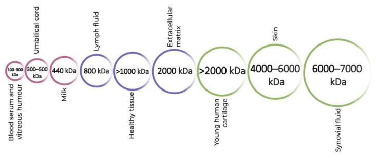
The nervous and immune systems go hand in hand in causing inflammation and pain. However, the two are not mutually exclusive. While some diseases cause inflammation, others are caused by it. Macrophages play an important role in modulating inflammation to trigger neuropathic pain. Hyaluronic acid (HA) is a naturally occurring glycosaminoglycan that has a well-known ability to bind with the cluster of differentiation 44 (CD44) receptor on classically activated M1 macrophages. Resolving inflammation by varying the molecular weight of HA is a debated concept. HA-based drug delivery nanosystems such as nanohydrogels and nanoemulsions, targeting macrophages can be used to relieve pain and inflammation by loading antinociceptive drugs and enhancing the effect of anti-inflammatory drugs. This review will discuss the ongoing research on HA-based drug delivery nanosystems regarding their antinociceptive and anti-inflammatory effects.
Keywords: CD44; anti-inflammatory; antinociceptive; hyaluronic acid; nanosystems.
Conflict of interest statement
The authors declare no conflict of interest.
Figures
Similar articles
-
CD44 Signaling Mediates High Molecular Weight Hyaluronan-Induced Antihyperalgesia.J Neurosci. 2018 Jan 10;38(2):308-321. doi: 10.1523/JNEUROSCI.2695-17.2017. Epub 2017 Nov 24. J Neurosci. 2018. PMID: 29175954 Free PMC article.
-
Hyaluronic acid based self-assembling nanosystems for CD44 target mediated siRNA delivery to solid tumors.Biomaterials. 2013 Apr;34(13):3489-502. doi: 10.1016/j.biomaterials.2013.01.077. Epub 2013 Feb 11. Biomaterials. 2013. PMID: 23410679 Free PMC article.
-
Lipid-based nanosystems for CD44 targeting in cancer treatment: recent significant advances, ongoing challenges and unmet needs.Nanomedicine (Lond). 2016 Jul;11(14):1865-87. doi: 10.2217/nnm-2016-5000. Epub 2016 Jul 7. Nanomedicine (Lond). 2016. PMID: 27389568 Review.
-
MicroRNA-223 Induced Repolarization of Peritoneal Macrophages Using CD44 Targeting Hyaluronic Acid Nanoparticles for Anti-Inflammatory Effects.PLoS One. 2016 May 5;11(5):e0152024. doi: 10.1371/journal.pone.0152024. eCollection 2016. PLoS One. 2016. PMID: 27148749 Free PMC article.
-
CD44 and its role in inflammation and inflammatory diseases.Inflamm Allergy Drug Targets. 2009 Jul;8(3):208-20. doi: 10.2174/187152809788680994. Inflamm Allergy Drug Targets. 2009. PMID: 19601881 Review.
Cited by
-
Hyaluronic Acid in Rheumatology.Pharmaceutics. 2023 Aug 30;15(9):2247. doi: 10.3390/pharmaceutics15092247. Pharmaceutics. 2023. PMID: 37765216 Free PMC article. Review.
-
Colon-Targeted Mucoadhesive PLGA Microspheres Loaded with Ramulus Mori Alkaloids for Enhanced Water-Soluble Drug Delivery in Ulcerative Colitis Treatment.Molecules. 2025 Apr 23;30(9):1878. doi: 10.3390/molecules30091878. Molecules. 2025. PMID: 40363686 Free PMC article.
-
Advances in hyaluronic acid hydrogel for meniscus repair.Front Bioeng Biotechnol. 2025 Jul 21;13:1639034. doi: 10.3389/fbioe.2025.1639034. eCollection 2025. Front Bioeng Biotechnol. 2025. PMID: 40761542 Free PMC article. Review.
-
Recent Advances in Nanoparticle and Nanocomposite-Based Photodynamic Therapy for Cervical Cancer: A Review.Cancers (Basel). 2025 Aug 4;17(15):2572. doi: 10.3390/cancers17152572. Cancers (Basel). 2025. PMID: 40805266 Free PMC article. Review.
-
CD44-targeting hyaluronic acid-selenium nanoparticles boost functional recovery following spinal cord injury.J Nanobiotechnology. 2024 Jan 23;22(1):37. doi: 10.1186/s12951-024-02302-0. J Nanobiotechnology. 2024. PMID: 38263204 Free PMC article.
References
-
- Calil I.L., Zarpelon A.C., Guerrero A.T.G., Alves-Filho J.C., Ferreira S.H., Cunha F.Q., Cunha T.M., Verri W.A., Jr. Lipopolysaccharide Induces Inflammatory Hyperalgesia Triggering a TLR4/MyD88-Dependent Cytokine Cascade in the Mice Paw. PLoS ONE. 2014;9:e90013. doi: 10.1371/journal.pone.0090013. - DOI - PMC - PubMed
Publication types
MeSH terms
Substances
Grants and funding
LinkOut - more resources
Full Text Sources
Miscellaneous
