. 2023 Apr 16;24(8):7340.
doi: 10.3390/ijms24087340.

Protective Effects of a Jellyfish-Derived Thioredoxin Fused with Cell-Penetrating Peptide TAT-PTD on H2O2-Induced Oxidative Damage

Affiliations

Protective Effects of a Jellyfish-Derived Thioredoxin Fused with Cell-Penetrating Peptide TAT-PTD on H2O2-Induced Oxidative Damage

Bo Wang et al. Int J Mol Sci. .

Abstract

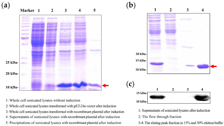
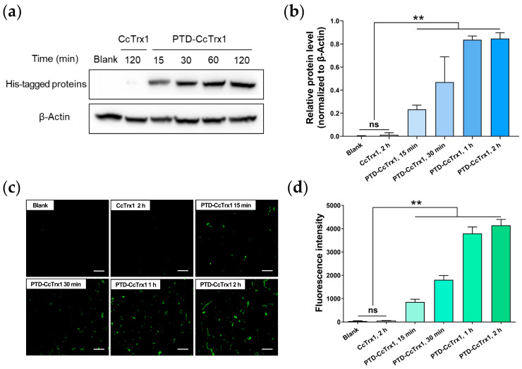
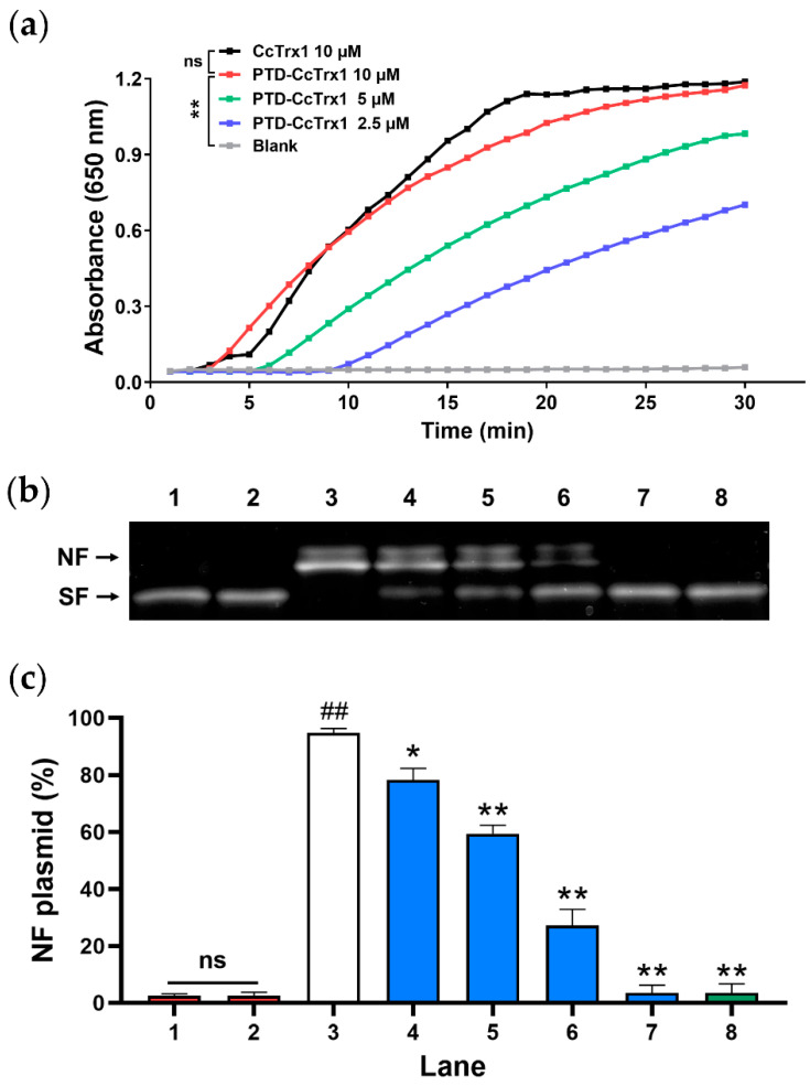
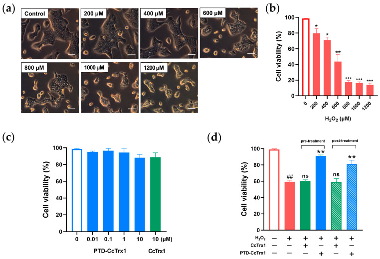
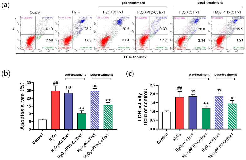
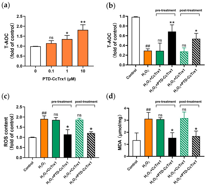
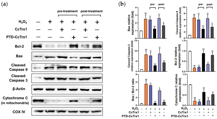
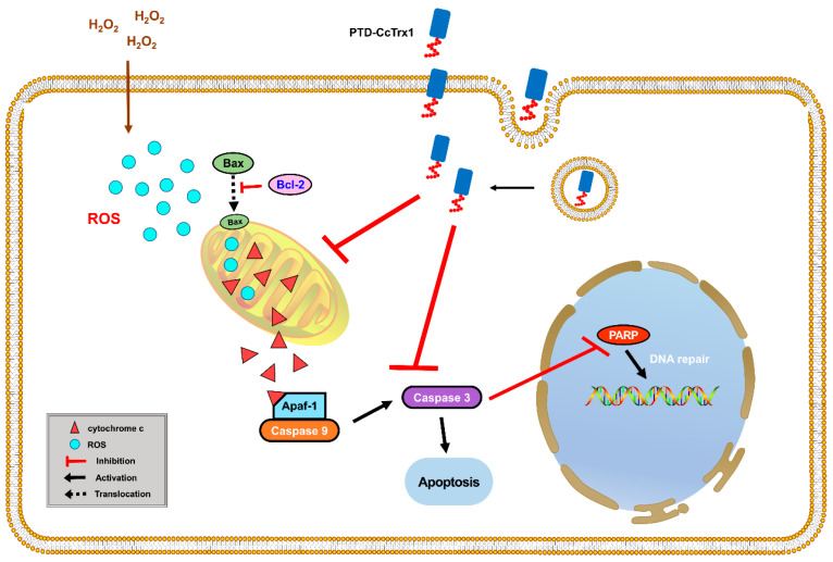
Thioredoxin (Trx) plays a critical role in maintaining redox balance in various cells and exhibits anti-oxidative, anti-apoptotic, and anti-inflammatory effects. However, whether exogenous Trx can inhibit intracellular oxidative damage has not been investigated. In previous study, we have identified a novel Trx from the jellyfish Cyanea capillata, named CcTrx1, and confirmed its antioxidant activities in vitro. Here, we obtained a recombinant protein, PTD-CcTrx1, which is a fusion of CcTrx1 and protein transduction domain (PTD) of HIV TAT protein. The transmembrane ability and antioxidant activities of PTD-CcTrx1, and its protective effects against H2O2-induced oxidative damage in HaCaT cells were also detected. Our results revealed that PTD-CcTrx1 exhibited specific transmembrane ability and antioxidant activities, and it could significantly attenuate the intracellular oxidative stress, inhibit H2O2-induced apoptosis, and protect HaCaT cells from oxidative damage. The present study provides critical evidence for application of PTD-CcTrx1 as a novel antioxidant to treat skin oxidative damage in the future.

Keywords: antioxidant; jellyfish; oxidative stress; protein transduction domain; thioredoxin.


Conflict of interest statement

The authors declare no conflict of interest.

Figures

Figure 1
Figure 1
Expression, purification, and identification of PTD-CcTrx1. Recombinant PTD-CcTrx1 plasmids were transformed into E. coli BL21, and then induced by 0.2 mM IPTG with shaking for 18 h at 37 °C. Cell pellets were collected, resuspended, and sonicated, and then centrifugated at 12,000× g for 30 min at 4 °C. The supernatants containing PTD-CcTrx1 were purified via the ÄKTA purifier system using a 5 mL HisTrap HP Chelating column. (a) SDS-PAGE analysis of the samples under different expression conditions. (b) SDS-PAGE analysis of the samples collected from different steps of purification. (c) Western blot analysis of the samples collected from different steps of purification using the anti-His-tag antibody. The position corresponding to PTD-CcTrx1 is indicated by the red arrow.
Figure 2
Figure 2
Transduction of PTD-CcTrx1 into HaCaT cells. First, HaCaT cells were incubated with 4 μM PTD-CcTrx1 for 15 min, 30 min, 1 h, 2 h, respectively. PBS and 4 μM CcTrx1 were used as a blank and negative control. Then, cells were rinsed with ice-cold PBS thoroughly, and the intracellular His-tagged proteins were detected by the anti-His-tag antibody via the Western blot assay and immunofluorescence assay. (a) Western blot analysis of intracellular His-tagged proteins using anti-His-tag antibody at different times. (b) Intracellular His-tagged proteins were quantified using densitometric analysis with β-Actin as the internal control. (c) Intracellular amount of His-tagged proteins at different times after incubation detected by immunofluorescence assay. Bar = 100 μm. (d) The fluorescence data were quantified by ImageJ software. Quantification data were obtained from three independent experiments, and the data are presented as means ± SD (n = 3) (ns, no significance, ** p < 0.01 vs. blank group).
Figure 3
Figure 3
Antioxidant activities of PTD-CcTrx1 in a cell-free system. (a) Disulfide reductase activity of PTD-CcTrx1. Insulin disulfide reduction activities of different concentrations of PTD-CcTrx1 and 10 μM CcTrx1 were measured in the presence of 2 mM DTT (ns, no significance, ** p < 0.01). (b,c) Effect of PTD-CcTrx1 on protecting supercoiled plasmid DNA against oxidative damage. Lane 1: pET-24a plasmid DNA alone; lane 2: pET-24a plasmid DNA, and 10 mM DTT; lane 3: pET-24a plasmid DNA, 10 mM DTT, and 35 μM FeCl3; lanes 4–7: pET-24a plasmid DNA, 10 mM DTT, 35 μM FeCl3, and different concentrations of PTD-CcTrx1 (1, 2, 4, 6 μM, respectively); lane 8: pET-24a plasmid DNA, 10 mM DTT, 35 μM FeCl3, and 6 μM CcTrx1. The bands corresponding to the nicked form (NF) and supercoiled form (SF) are indicated on the left side. Quantification data were obtained from three independent experiments, and the data are presented as means ± SD (n = 3) (ns, no significance, ## p < 0.01 vs. lane 1; * p < 0.05, ** p < 0.01 vs. lane 3).
Figure 4
Figure 4
Protective effects of PTD-CcTrx1 against H2O2-induced cytotoxicity in HaCaT cells. (a) Representative morphological changes of HaCaT cells, which were treated with different concentrations of H2O2 and observed with a phase contrast microscope. Bar = 100 μm. (b) HaCaT cells were treated with increasing concentrations of H2O2 (0, 200, 400, 600, 800, 1000, 1200 μM) for 4 h (* p < 0.05, ** p < 0.01, *** p < 0.001 vs. control group). (c) HaCaT cells were treated with increasing concentrations of PTD-CcTrx1 (0, 0.01, 0.1, 1, 10 μM) and 10 μM CcTrx1 for 12 h. (d) HaCaT cells were treated with 10 μM PTD-CcTrx1 and 10 μM CcTrx1 before or after the exposure to 400 μM H2O2. All treatments were performed in triplicate individually, and the data are presented as means ± SD (n = 3) (## p < 0.01 vs. control group; ns, no significance, ** p < 0.01 vs. H2O2 alone group).
Figure 5
Figure 5
Protective effects of PTD-CcTrx1 against H2O2-induced apoptosis and necrosis in HaCaT cells. First, HaCaT cells were treated with 10 μM PTD-CcTrx1 and 10 μM CcTrx1 before or after the exposure to 400 μM H2O2. After treatment, the cells were resuspended in the binding buffer of the apoptosis analysis kit and stained with Annexin V-FITC/PI, and then analyzed by flow cytometry. (a) PTD-CcTrx1 significantly inhibited H2O2-induced apoptosis of HaCaT cells. (b) Histogram of the apoptosis rate in different groups. (c) LDH activity of cell supernatants in different groups. All treatments were performed in triplicate individually, with values expressed as mean ± SD (## p < 0.01 vs. control; ns, no significance, * p < 0.05, ** p < 0.01 vs. H2O2 alone group).
Figure 6
Figure 6
Attenuation of H2O2-mediated oxidative stress in HaCaT cells by PTD-CcTrx1. (a) The total antioxidant capacity (T-AOC) in HaCaT cells after treatment with different concentrations of PTD-CcTrx1 for 2 h (ns, no significance, * p < 0.05, ** p < 0.01 vs. control group). (b) PTD-CcTrx1 attenuated the decrease of T-AOC induced by H2O2. HaCaT cells were treated with 10 μM CcTrx1 and 10 μM PTD-CcTrx1 before or after the exposure to 400 μM H2O2. Afterward, the cells were rinsed with ice-cold PBS and treated with 80 μL of cell lysis buffer. Then, cell lysates were scraped thoroughly for centrifugation, and the supernatants were collected to detect the T-AOC. (c) ROS increase induced by H2O2 was attenuated by PTD-CcTrx1. After treatment, HaCaT cells were rinsed with PBS before undergoing DCFH-DA staining and then detected under a fluorescence microplate reader. (d) Analysis of MDA generation and lipid peroxidation. After treatment, cell lysates were scraped thoroughly and the supernatants were collected to quantify the MDA level. All treatments were performed in triplicate individually, with values expressed as mean ± SD (## p < 0.01 vs. control; ns, no significance, * p < 0.05, ** p < 0.01 vs. H2O2 alone group).
Figure 7
Figure 7
PTD-CcTrx1 inhibited the intrinsic apoptotic pathway in H2O2-treated HaCaT cells. First, HaCaT cells were treated with 10 μM CcTrx1 and 10 μM PTD-CcTrx1 before or after the exposure to 400 μM H2O2. The cells were washed with ice-cold PBS, and cytoplasmic and mitochondrial proteins were extracted for the Western blot assay. (a) Western blot analysis of Bax, Bcl-2, cleaved-caspase 3, cleaved-caspase 9, and cytochrome c (in mitochondria) of each group. β-Actin and COX IV were set as internal references, respectively. (b) Relative contents of the proteins were calibrated by setting the control group as one. All treatments were performed in triplicate individually, and the data are presented as means ± SD (n = 3) (## p < 0.01 vs. control; ns, no significance, * p < 0.05, ** p < 0.01 vs. H2O2 alone group).
Figure 8
Figure 8
Possible mechanisms of the protective effect of PTD-CcTrx1 against H2O2-induced apoptosis in HaCaT cells. H2O2 induced the accumulation of intracellular ROS, which induced apoptosis by promoting the translocation of Bax and cytochrome c and then activating caspase 9 and caspase 3. PTD-CcTrx1 could penetrate the cell membrane and protect against H2O2-induced apoptosis by downregulating the Bax/Bcl-2 ratio, inhibiting the translocation of cytochrome c, and suppressing the activation of caspase 9 and caspase 3.

Similar articles

Cited by

References

    1. Mohania D., Chandel S., Kumar P., Verma V., Digvijay K., Tripathi D., Choudhury K., Mitten S.K., Shah D. Ultraviolet radiations: Skin defense-damage mechanism. Adv. Exp. Med. Biol. 2017;996:71–87. - PubMed
    1. D’Orazio J., Jarrett S., Amaro-Ortiz A., Scott T. UV radiation and the skin. Int. J. Mol. Sci. 2013;14:12222–12248. doi: 10.3390/ijms140612222. - DOI - PMC - PubMed
    1. Ahn K. The role of air pollutants in atopic dermatitis. J. Allergy Clin. Immunol. 2014;134:993–999. doi: 10.1016/j.jaci.2014.09.023. - DOI - PubMed
    1. Celebi Sözener Z., Cevhertas L., Nadeau K., Akdis M., Akdis C.A. Environmental factors in epithelial barrier dysfunction. J. Allergy Clin. Immunol. 2020;145:1517–1528. doi: 10.1016/j.jaci.2020.04.024. - DOI - PubMed
    1. Gu Y., Han J., Jiang C., Zhang Y. Biomarkers, oxidative stress and autophagy in skin aging. Ageing Res. Rev. 2020;59:101036. doi: 10.1016/j.arr.2020.101036. - DOI - PubMed

MeSH terms

LinkOut - more resources