

Visual Evoked Potentials for the Detection of Diabetic Retinal Neuropathy
- PMID: 37108524
- PMCID: PMC10138821
- DOI: 10.3390/ijms24087361
Visual Evoked Potentials for the Detection of Diabetic Retinal Neuropathy
Abstract
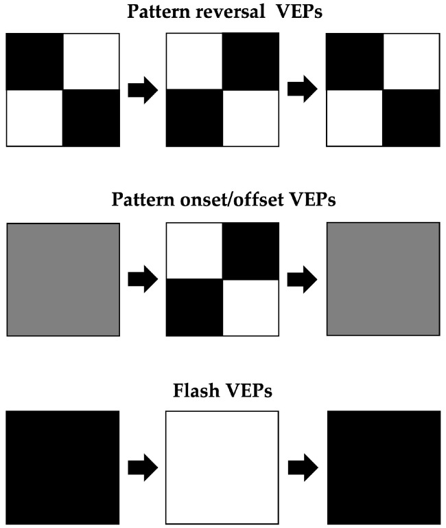
Visual evoked potentials (VEP) are visually evoked signals that extract electroencephalographic activity in the visual cortex that can detect retinal ganglion cells, optic nerves, chiasmal and retrochiasmal dysfunction, including optic radiations, and the occipital cortex. Because diabetes causes diabetic retinopathy due to microangiopathy and neuropathy due to metabolic abnormalities and intraneural blood flow disorders, assessment of diabetic visual pathway impairment using VEP has been attempted. In this review, evidence on the attempts to assess the visual pathway dysfunction due to abnormal blood glucose levels using VEP is presented. Previous studies have provided significant evidence that VEP can functionally detect antecedent neuropathy before fundus examination. The detailed correlations between VEP waveforms and disease duration, HbA1c, glycemic control, and short-term increases and decreases in blood glucose levels are evaluated. VEP may be useful for predicting postoperative prognosis and evaluating visual function before surgery for diabetic retinopathy. Further controlled studies with larger cohorts are needed to establish a more detailed relationship between diabetes mellitus and VEP.
Keywords: diabetes mellitus; diabetic retinopathy; visual evoked potential.
Conflict of interest statement
The author declare no conflict of interest.
Figures
Similar articles
-
Utility of Visual Evoked Potentials (VEPs) study in the evaluation of visual pathway dysfunction in diabetics without retinopathy: correlations with diabetic peripheral neuropathy and other clinical findings.Rom J Ophthalmol. 2024 Apr-Jun;68(2):114-121. doi: 10.22336/rjo.2024.22. Rom J Ophthalmol. 2024. PMID: 39006331 Free PMC article.
-
Electrophysiological changes in optic neuropathy of streptozotocin induced diabetic rats.J Med Life. 2013 Sep 15;6(3):340-8. Epub 2013 Sep 25. J Med Life. 2013. PMID: 24155786 Free PMC article.
-
Evaluation of visual pathways using visual evoked potential in patients with diabetic retinopathy.Rom J Ophthalmol. 2019 Oct-Dec;63(4):367-371. Rom J Ophthalmol. 2019. PMID: 31915735 Free PMC article.
-
Role of Electrophysiology in the Early Diagnosis and Follow-Up of Diabetic Retinopathy.J Diabetes Res. 2015;2015:319692. doi: 10.1155/2015/319692. Epub 2015 May 5. J Diabetes Res. 2015. PMID: 26075282 Free PMC article. Review.
-
Evoked potentials in diabetes mellitus.Clin Neurosci. 1997;4(6):374-9. Clin Neurosci. 1997. PMID: 9358983 Review.
Cited by
-
Retinal Function in Long-Term Type 1 Diabetes without Retinopathy: Insights from Pattern Electroretinogram and Pattern Visual Evoked Potentials Assessments.Diagnostics (Basel). 2024 Feb 25;14(5):492. doi: 10.3390/diagnostics14050492. Diagnostics (Basel). 2024. PMID: 38472964 Free PMC article.
-
Angiogenic and Fibrogenic Dual-effect of Gremlin1 on Proliferative Diabetic Retinopathy.Int J Biol Sci. 2024 Jan 12;20(3):897-915. doi: 10.7150/ijbs.85735. eCollection 2024. Int J Biol Sci. 2024. PMID: 38250154 Free PMC article.
-
Assessment of Visual Evoked Potential to Detect Visual Pathway Dysfunction in Gestational Diabetes Mellitus: A Longitudinal Case-Control Study With Postpartum Follow-up.Cureus. 2023 Nov 29;15(11):e49619. doi: 10.7759/cureus.49619. eCollection 2023 Nov. Cureus. 2023. PMID: 38161906 Free PMC article.
-
Bright light therapy in Parkinson's disease: a pilot study on visual pathway improvements.BMC Psychiatry. 2025 May 12;25(1):476. doi: 10.1186/s12888-025-06915-z. BMC Psychiatry. 2025. PMID: 40355852 Free PMC article. Clinical Trial.
-
Clinical observations and mechanistic insights of traditional Chinese medicine in the management of diabetic retinopathy.Pharm Biol. 2024 Dec;62(1):529-543. doi: 10.1080/13880209.2024.2369292. Epub 2024 Jun 26. Pharm Biol. 2024. PMID: 38921697 Free PMC article. Review.
References
-
- Wong T.Y., Sun J., Kawasaki R., Ruamviboonsuk P., Gupta N., Lansingh V.C., Maia M., Mathenge W., Moreker S., Muqit M.M.K., et al. Guidelines on Diabetic Eye Care: The International Council of Ophthalmology Recommendations for Screening, Follow-up, Referral, and Treatment Based on Resource Settings. Ophthalmology. 2018;125:1608–1622. doi: 10.1016/j.ophtha.2018.04.007. - DOI - PubMed
Publication types
MeSH terms
Substances
LinkOut - more resources
Full Text Sources
Medical
