

Localized Cutaneous Nodular Amyloidosis: A Specific Cutaneous Manifestation of Sjögren's Syndrome
- PMID: 37108553
- PMCID: PMC10139233
- DOI: 10.3390/ijms24087378
Localized Cutaneous Nodular Amyloidosis: A Specific Cutaneous Manifestation of Sjögren's Syndrome
Abstract
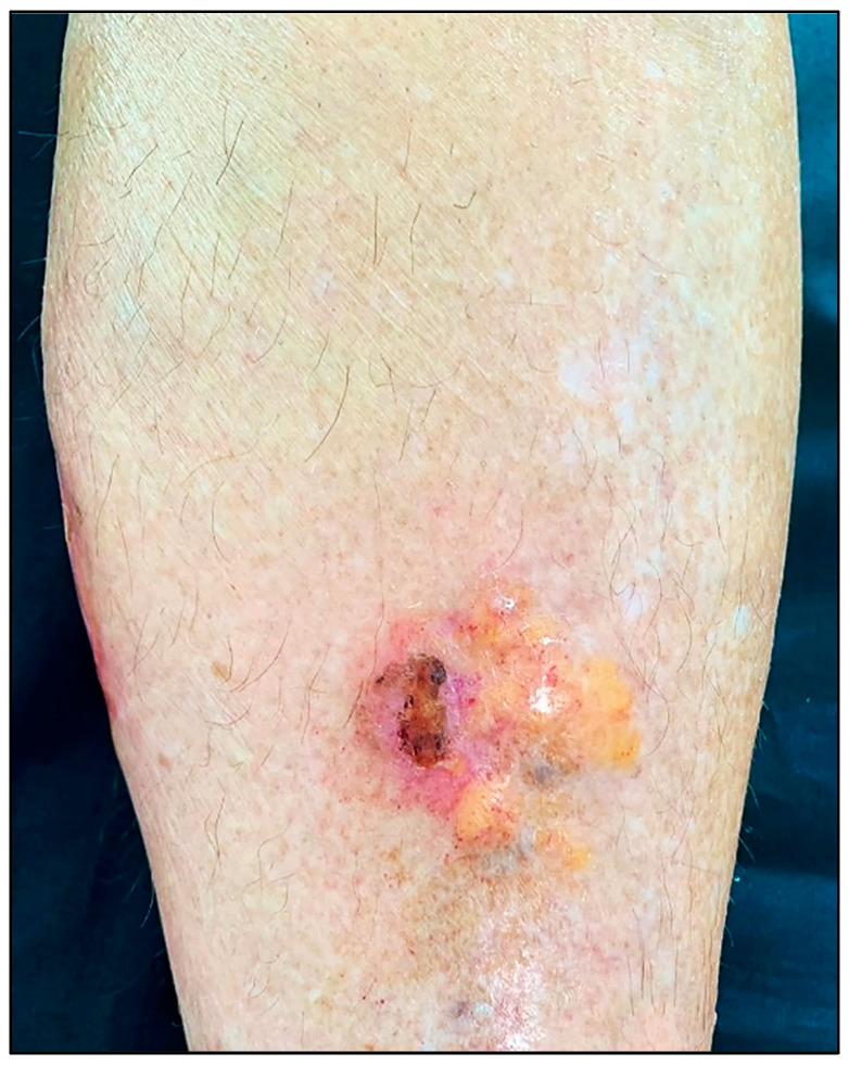
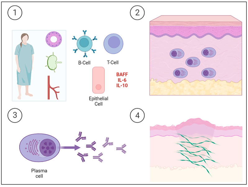
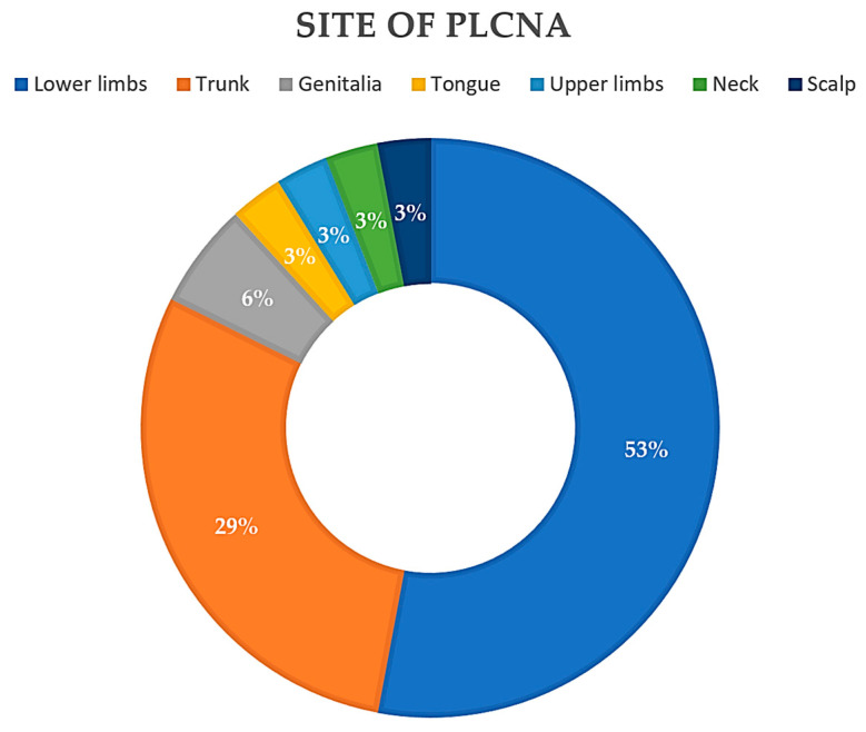
Primary localized cutaneous nodular amyloidosis (PLCNA) is a rare condition attributed to plasma cell proliferation and the deposition of immunoglobulin light chains in the skin without association with systemic amyloidosis or hematological dyscrasias. It is not uncommon for patients diagnosed with PLCNA to also suffer from other auto-immune connective tissue diseases, with Sjögren's syndrome (SjS) showing the strongest association. This article provides a literature review and descriptive analysis to better understand the unique relationship between these two entities. To date, 34 patients with PLCNA and SjS have been reported in a total of 26 articles. The co-existence of PLCNA and SjS has been reported, especially in female patients in their seventh decade of life with nodular lesions on the trunk and/or lower extremities. Acral and facial localization, which is a typical localization of PLCNA in the absence of SjS, seems to be much more unusual in patients with associated SjS.
Keywords: Sjögren’s Syndrome; auto-immune connective tissue disorders; localized cutaneous nodular amyloidosis; plasma cells.
Conflict of interest statement
The authors have no conflict of interest to declare.
Figures
Similar articles
-
Localized Cutaneous Nodular Amyloidosis in a Patient with Sjögren's Syndrome.Int J Mol Sci. 2023 May 28;24(11):9409. doi: 10.3390/ijms24119409. Int J Mol Sci. 2023. PMID: 37298361 Free PMC article.
-
Primary localized cutaneous nodular amyloidosis in a patient with Sjögren's syndrome: a review of the literature.J Dermatol. 2005 Feb;32(2):120-3. doi: 10.1111/j.1346-8138.2005.tb00728.x. J Dermatol. 2005. PMID: 15906542 Review.
-
Characterization of the amyloid fibril from primary localized cutaneous nodular amyloidosis associated with Sjögren's syndrome.Dermatology. 1994;189(2):125-8. doi: 10.1159/000246814. Dermatology. 1994. PMID: 8075437 Review.
-
Primary Localized Cutaneous Nodular Amyloidosis and Limited Cutaneous Systemic Sclerosis: Additional Cases with Dermatoscopic and Histopathological Correlation of Amyloid Deposition.Dermatopathology (Basel). 2021 Jul 2;8(3):229-235. doi: 10.3390/dermatopathology8030028. Dermatopathology (Basel). 2021. PMID: 34287266 Free PMC article.
-
[Nodular cutaneous amyloidosis associated with Sjögren's syndrome].Ann Dermatol Venereol. 2013 May;140(5):378-81. doi: 10.1016/j.annder.2013.02.014. Epub 2013 Mar 29. Ann Dermatol Venereol. 2013. PMID: 23663711 French.
Cited by
-
A 71-year-old Woman with CREST Syndrome and Multiple Waxy Facial Papules and Plaques: A Quiz.Acta Derm Venereol. 2024 Jun 8;104:adv40419. doi: 10.2340/actadv.v104.40419. Acta Derm Venereol. 2024. PMID: 38850086 Free PMC article. No abstract available.
-
Histopathological Insights into Primary Localized Cutaneous Amyloidosis: A Case Series.Cureus. 2025 Feb 24;17(2):e79603. doi: 10.7759/cureus.79603. eCollection 2025 Feb. Cureus. 2025. PMID: 40151750 Free PMC article.
-
Associations between metabolic disorders and Sjögren's disease.Jpn Dent Sci Rev. 2024 Dec;60:232-238. doi: 10.1016/j.jdsr.2024.06.002. Epub 2024 Oct 21. Jpn Dent Sci Rev. 2024. PMID: 39502167 Free PMC article. Review.
-
Primary Localized Labial Amyloidosis Associated with Sjögren Syndrome.Head Neck Pathol. 2024 Aug 13;18(1):74. doi: 10.1007/s12105-024-01679-6. Head Neck Pathol. 2024. PMID: 39136797 Free PMC article. No abstract available.
References
-
- Meijer J.M., Schonland S.O., Palladini G., Merlini G., Hegenbart U., Ciocca O., Perfetti V., Leijsma M.K., Bootsma H., Hazenberg B.P. Sjögren’s syndrome and localized nodular cutaneous amyloidosis: Coincidence or a distinct clinical entity? Arthritis Rheum. 2008;58:1992–1999. doi: 10.1002/art.23617. - DOI - PubMed
Publication types
MeSH terms
Substances
LinkOut - more resources
Full Text Sources
Medical
Molecular Biology Databases
