. 2023 Apr 18;24(8):7439.
doi: 10.3390/ijms24087439.

Preclinical Evaluation of TB/FLU-04L-An Intranasal Influenza Vector-Based Boost Vaccine against Tuberculosis

Affiliations

Preclinical Evaluation of TB/FLU-04L-An Intranasal Influenza Vector-Based Boost Vaccine against Tuberculosis

Anna-Polina Shurygina et al. Int J Mol Sci. .

Abstract

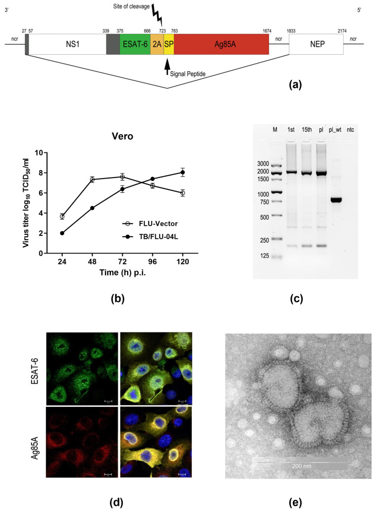
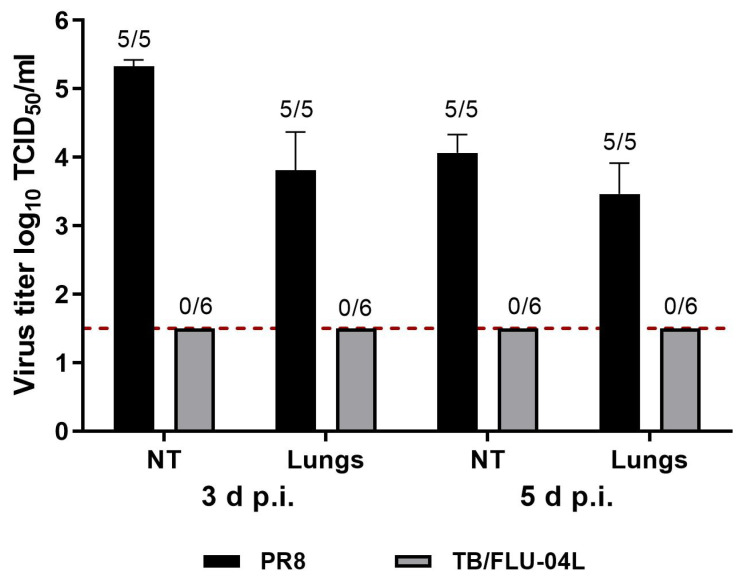
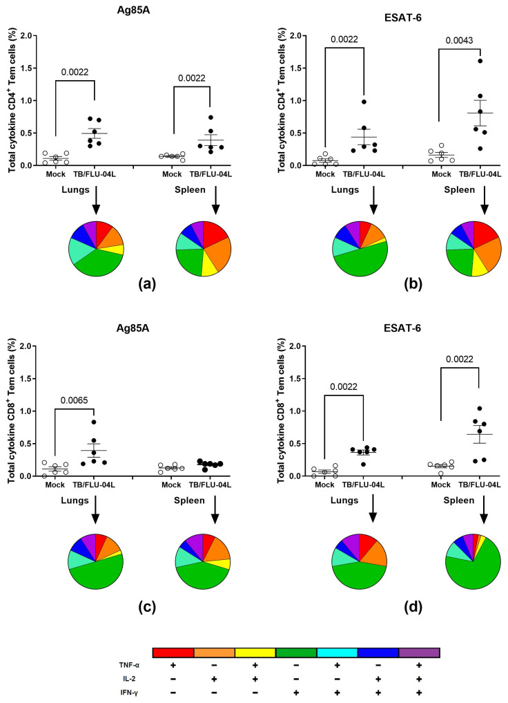
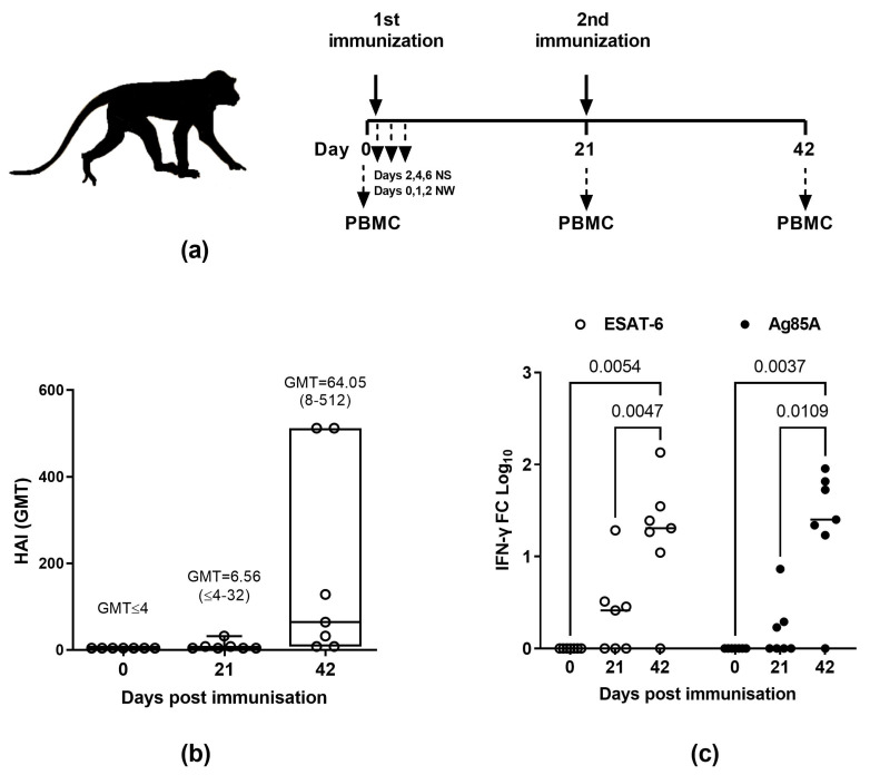
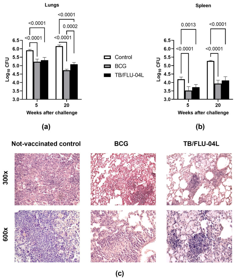
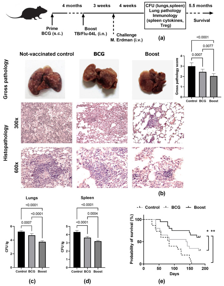
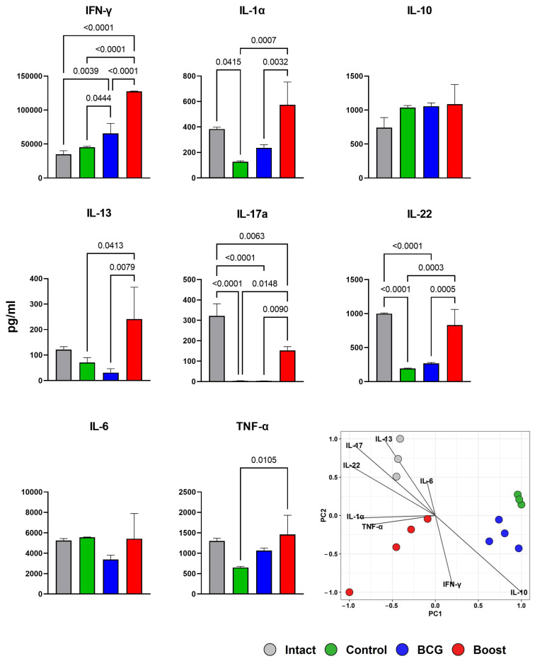
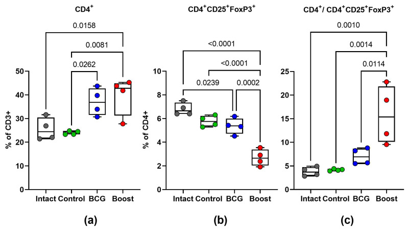
Tuberculosis is a major global threat to human health. Since the widely used BCG vaccine is poorly effective in adults, there is a demand for the development of a new type of boost tuberculosis vaccine. We designed a novel intranasal tuberculosis vaccine candidate, TB/FLU-04L, which is based on an attenuated influenza A virus vector encoding two mycobacterium antigens, Ag85A and ESAT-6. As tuberculosis is an airborne disease, the ability to induce mucosal immunity is one of the potential advantages of influenza vectors. Sequences of ESAT-6 and Ag85A antigens were inserted into the NS1 open reading frame of the influenza A virus to replace the deleted carboxyl part of the NS1 protein. The vector expressing chimeric NS1 protein appeared to be genetically stable and replication-deficient in mice and non-human primates. Intranasal immunization of C57BL/6 mice or cynomolgus macaques with the TB/FLU-04L vaccine candidate induced Mtb-specific Th1 immune response. Single TB/FLU-04L immunization in mice showed commensurate levels of protection in comparison to BCG and significantly increased the protective effect of BCG when applied in a "prime-boost" scheme. Our findings show that intranasal immunization with the TB/FLU-04L vaccine, which carries two mycobacterium antigens, is safe, and induces a protective immune response against virulent M. tuberculosis.

Keywords: Ag85A; ESAT-6; M. tuberculosis vaccine; influenza vector; mucosal immunization.


Conflict of interest statement

The authors declare no conflict of interest. The funders had no role in the design of the study; in the collection, analyses, or interpretation of data; in the writing of the manuscript or in the decision to publish the results.

Figures

Figure 1
Figure 1
Construction and molecular characterization of the vaccine strain. (a) Schematic representation of the recombinant NS gene segment expressing antigens ESAT-6 and Ag85A of M. tuberculosis. The gray box (27–57) represents 12 aa that are shared between the NS1 and NEP proteins; the gray box (339–375) represents nucleotides encoding a random 12aa segment, fused to nucleotides encoding the ESAT-6 (green box) antigen followed by an autoproteolytic 2A cleavage site (orange box). The yellow box represents the nucleotide sequence of the modified mouse IgK-derived signal peptide (SP) followed by the Ag85A sequence (red box); ncr is a non-coding region. Nucleotide positions are indicated at the top. (b) Growth curve of TB/FLU-04L in Vero cells in comparison to an empty vector (FLU-vector). (c) Genetic stability of the chimeric NS1 gene. RT-PCR analysis of the chimeric NS116_ESAT-6_Ag85A segment in Flu/ESAT-6_Ag85A virus after 2 (P2) and 15 (P15) passages in Vero cells. Viral RNA was isolated from the supernatant of infected cells at 72 h p.i., and One-Step qRT-PCR was performed. The control used was pHW plasmid encoding NS116_ESAT-6_Ag85A. M, Marker; pl, plasmid DNA; wt pl, pHW plasmid encoding NS1 gene segment from influenza A/PR8/34 wt virus. (d) Expression of ESAT-6, Ag85A, and NP proteins in infected Vero cells. Cells were infected with the Flu/ESAT-6_Ag85A virus (P5) at a multiplicity of infection (m.o.i.) of 2, then fixed and stained at 16 h p.i. using anti-ESAT-6 (green) or anti-Ag85A (red), anti-NP (yellow) antibodies, and DAPI (blue). Scale bars (white) correspond to 10 μm (e) Electron microphotograph of TB/FLU-04L viral particles following purification.
Figure 2
Figure 2
Virus shedding. Groups of ten or twelve 7–8 weeks old female C57bl/6 mice were inoculated with 0.01 mL of either TB/FLU-04L in a dose 6.0 log10 TCID50/animal or A/PR/8/34 in a dose 5.0 log10 TCID50/animal under slight ether anesthesia. Viral loads in 10% suspensions of nasal turbines and lungs were determined on days 3 and 5 post-inoculation in Vero cells. The virus titers are expressed as the mean log10 TCID50/mL ± SEM from 5–6 animals. The limit of virus detection was 1.5 log10 TCID50/mL (red dotted line). Tissues, where no virus was detected, were assigned the value of 1.5 log10 TCID50/mL for the calculation of the mean titer. The numbers placed above the bars reflect the ratio of animals with virus shedding to the total number of animals in the group. Mice with a lung virus load <1.5 log10 TCID50/mL were classified as not infected.
Figure 3
Figure 3
Antigen-specific immune response to TB/FLU-04L. Total frequency (%) of Ag85A (a,c) and ESAT-6-specific (b,d) cytokine-secreting CD4+ (a,b) and CD8+ (c,d) Tem cells were measured following a single TB/FLU-04L vaccination. Groups of six 7–8 weeks old female C57bl/6 mice were inoculated with 0.01 mL of DPBS (Mock) or TB/FLU-04L in a dose of 6.0 log10 TCID50/animal under slight ether anesthesia. Three weeks after the vaccination, single-cell suspensions prepared from the lungs and spleens of vaccinated mice were stimulated by the ESAT-6 and Ag85A recombinant proteins in vitro. The frequencies of IFN-γ-, TNF-α-, and IL-2-producing CD4+ and CD8+ Tem cells were measured by flow cytometry (ICS). Background staining from cells stimulated with medium alone has been subtracted. The data were considered statistically significant when p < 0.05 in the Mann–Whitney test.
Figure 4
Figure 4
Immunogenicity of TB/FLU-04L in Macaca fascicularis. (a) Study timeline and sampling schedule. (b) Vector-specific antibody response in HAI and (c) antigen-specific IFNγ response (fold change [FC] log10) after the i.n. vaccination with TB/FLU-04L: seven adult male Macaca fascicularis were immunized twice with TB/FLU-04 (7.5 log10TCID50/animal), with a three-week interval between vaccinations. Blood samples for HAI assay and PBMCs were collected before each vaccination (day 0, day 21) and three weeks after the second vaccination (day 42). To assess the antigen-specific IFNγ response, PBMCs were stimulated in vitro by M. tuberculosis antigens (ESAT-6 or Ag85A 5μg/mL) for 72 h. Medium alone or Concanavalin A were used as a negative or positive control, respectively. Following incubation, cell supernatants were collected and IFNγ levels were determined by using the BD OptEIA™ Monkey ELISA Set. The data were considered statistically significant when p < 0.05 in the two-way ANOVA followed by Tukey’s multiple comparison test.
Figure 5
Figure 5
Protective immunity against virulent M. tuberculosis intravenous challenge in mice following intranasal vaccination with TB/FLU-04L. C57BL/6 mice received saline i.n. (naive), or 1 × 10⁵ BCG s.c., or TB/FLU-04L i.n. twice with three-week intervals. Three weeks after the second vaccination, mice were challenged by an i.v. injection of 106 CFU of virulent M. tuberculosis Erdman strain. Graphs show means and SDs of CFU of M. tuberculosis in the lung (a) and spleen (b) 5 and 20 weeks after the challenge for groups of five mice that were either non-vaccinated control (white bars), BCG vaccinated (gray bars), or TB/FLU-04L vaccinated (black bars). The data were considered statistically significant at p < 0.05 in the mixed-model analysis with Tukey’s multiple comparisons test. (c) Development of lung pathology in three experimental groups of C57BL/6 mice five weeks after the i.v. challenge with M. tuberculosis Erdman (n = 10 per group). The photographs represent pathological lung tissue samples from the non-vaccinated control and mice vaccinated with BCG or TB/FLU-04L. H&E staining, magnification 300× (upper row), and 600× (lower row).
Figure 6
Figure 6
Protective efficacy of the BCG prime with TB/FLU-04L boost immunization against the i.v. M. tuberculosis challenge in C57BL/6 mice. (a) Study timeline and sampling schedule. C57BL/6 mice (40 animals per group) were immunized with a single s.c. dose of BCG (105 CFU; BCG group) or with one dose of BCG administered s.c. followed by one intranasal immunization with the influenza vector expressing ESAT-6 and Ag85A proteins (106 TCID50, BCG prime/TB/FLU-04 boost group) four months apart. The control group was immunized with PBS only at the time of the prime and boost immunizations (non-vaccinated control group). Three weeks after the boost immunization, mice were challenged intravenously (i.v.) with the virulent Erdman strain of M. tuberculosis (1 × 10⁶ CFU). Four weeks after the challenge, the level of protection was measured by enumerating bacterial loads (CFU) in the lungs and spleens (6 animals per group) and evaluating gross pathological and histopathological changes in the lungs (10 animals per group). Cytokine secretion by spleen cells was measured at the time of sacrifice (4 mice per group). The survival of vaccinated and non-vaccinated C57BL/6 mice (15–20 animals per group) following the i.v. infection with M. tuberculosis Erdman was monitored over the 184-day observation period. (b) Tuberculosis lesions in the lungs of infected mice (n = 12 per group). Representative photographs of lungs, gross pathology scores, and histopathological appearances of the lung tissues stained with H&E. Magnification 300× (upper row) and 600× (lower row). (c,d) Bacterial growth in the lungs and spleens of vaccinated and control mice. The data were considered statistically significant when p < 0.05, as determined by the one-way ANOVA with Tukey’s multiple comparisons test. (e) The survival of C57BL/6 mice for 184 days post-infection. The significance of the differences between survival times was evaluated using the Log-rank test (* = p < 0.01, ** = p < 0.001).
Figure 7
Figure 7
Spleen cell cytokine production in the BCG-vaccinated mice with or without TB/FLU-04L boost after M. tuberculosis infection. Four weeks after the i.v. challenge, spleen cells from intact (gray bars), control non-vaccinated mice (green bars), BCG-vaccinated mice (blue bars), or BCG-vaccinated/TB/FLU-04L-boosted mice (red bars) were cultured in the presence of medium only, ConA (2.5 μg/mL), or BCG (5 μg/mL). Levels of cytokines in the culture supernatants were measured after 72 h by using the multiplex kit (Biolegend). Values are expressed in pg/mL and represent group medians ± SEMs of samples tested in duplicates. The data were considered statistically significant when p < 0.05 in one-way ANOVA with Tukey’s multiple comparisons test. Additionally, visualization of differences in cytokine response between the groups was performed using the Principal Component Analysis (PCA).
Figure 8
Figure 8
Treg-cell frequency during the M. tuberculosis challenge. Four weeks after the i.v. challenge, relative amounts of CD4+ T cells (a), CD4+CD25+FoxP3+ Treg cells (b), and their ratios (c) were evaluated in spleen cells collected from intact mice (gray), non-vaccinated control mice (blue), BCG-vaccinated mice (green), or BCG-vaccinated/TB/FLU-04L-boosted mice (red). The data were considered statistically significant when p < 0.05 in one-way ANOVA with Tukey’s multiple comparisons test.

Similar articles

Cited by

References

    1. World Health Organization . Global Tuberculosis Report 2022. Volume 4. IGO; Geneva, Switzerland: 2022.
    1. Lipman M., McQuaid C.F., Abubakar I., Khan M., Kranzer K., McHugh T.D., Padmapriyadarsini C., Rangaka M.X., Stoker N. The Impact of COVID-19 on Global Tuberculosis Control. Indian J. Med. Res. 2021;153:404–408. doi: 10.4103/ijmr.IJMR_326_21. - DOI - PMC - PubMed
    1. Kumar P. A Perspective on the Success and Failure of BCG. Front. Immunol. 2021;12:778028. doi: 10.3389/fimmu.2021.778028. - DOI - PMC - PubMed
    1. Lavelle E.C., Ward R.W. Mucosal Vaccines—Fortifying the Frontiers. Nat. Rev. Immunol. 2022;22:236–250. doi: 10.1038/s41577-021-00583-2. - DOI - PMC - PubMed
    1. Ogongo P., Porterfield J.Z., Leslie A. Lung Tissue Resident Memory T-Cells in the Immune Response to Mycobacterium Tuberculosis. Front. Immunol. 2019;10:992. doi: 10.3389/fimmu.2019.00992. - DOI - PMC - PubMed