Review
. 2023 Apr 19;24(8):7481.
doi: 10.3390/ijms24087481.

Retinitis Pigmentosa: Current Clinical Management and Emerging Therapies

Affiliations
Review

Retinitis Pigmentosa: Current Clinical Management and Emerging Therapies

Xuan-Thanh-An Nguyen et al. Int J Mol Sci. .

Abstract

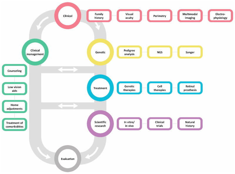
Retinitis pigmentosa (RP) comprises a group of inherited retinal dystrophies characterized by the degeneration of rod photoreceptors, followed by the degeneration of cone photoreceptors. As a result of photoreceptor degeneration, affected individuals experience gradual loss of visual function, with primary symptoms of progressive nyctalopia, constricted visual fields and, ultimately, central vision loss. The onset, severity and clinical course of RP shows great variability and unpredictability, with most patients already experiencing some degree of visual disability in childhood. While RP is currently untreatable for the majority of patients, significant efforts have been made in the development of genetic therapies, which offer new hope for treatment for patients affected by inherited retinal dystrophies. In this exciting era of emerging gene therapies, it remains imperative to continue supporting patients with RP using all available options to manage their condition. Patients with RP experience a wide variety of physical, mental and social-emotional difficulties during their lifetime, of which some require timely intervention. This review aims to familiarize readers with clinical management options that are currently available for patients with RP.

Keywords: clinical management; gene therapy; genetic counseling; genetics; low vision; low-vision rehabilitation; retinitis pigmentosa.


Conflict of interest statement

The authors declare no conflict of interest.

Figures

Figure 1
Figure 1
Venn diagram of currently identified genes associated with retinitis pigmentosa (RP) and their genetic overlap with other inherited retinal dystrophies. For example, variants in the RHO gene can manifest in either RP or congenital stationary night-blindness phenotypes. All genes included are registered in the Online Mendelian Inheritance in Man (OMIM) database and follow the up-to-date symbols of the HUGO Gene Nomenclature Committee (HGNC). Genes that are associated with syndromic forms of RP are marked with an asterisk (*). ACHM = achromatopsia; CO(R)D = cone(-rod) dystrophy; CSNB = congenital stationary night blindness; LCA = Leber Congenital Amaurosis; MD = macular dystrophy.
Figure 2
Figure 2
Illustrative example of typical visual field progression in a patient with retinitis pigmentosa using kinetic perimetry. Visual fields can be within normal limits in early stages of disease (I), although visual field defects may already be present but not detectable within the used target stimulus. With time, constriction of the visual fields occurs, with defects typically being symmetric and expanding more rapidly outwards and slower inwards (II,III). Ultimately, a small central remnant of visual field may remain in end-stage retinitis pigmentosa, which is commonly experienced and known as ‘tunnel vision’ (IV). Note that the clinical course of visual field loss varies between individuals and may follow a progression pattern that is different from this illustration.
Figure 3
Figure 3
Example full-field electroretinography recordings in a healthy patient and in patients with different disease stages of retinitis pigmentosa. Different stimuli are used to establish the diagnosis of retinitis pigmentosa, which is based on the guidelines of the International Society for Clinical Electrophysiology of Vision (ISCEV). In patients with advanced stages of diseases, rod-driven responses are severely diminished or even absent, whereas residual cone-driven responses may still remain.
Figure 4
Figure 4
Multimodal imaging in three patients with retinitis pigmentosa (RP). (AC): Multimodal imaging in a patient with RP caused by a variant in the RHO gene showing the clinical hallmarks of RP, including attenuated vessels and bone-spicule-like hyperpigmentation in the (mid)peripheral retina (A). On autofluorescence imaging, a small hyperfluorescent ring is observed in the macula (B). Spectral-domain optical coherence imaging shows a relatively intact central retina with loss of the outer retinal layers (i.e., ellipsoid zone and external limiting membrane) outside this area (C). (DF): Multimodal imaging in a different patient with RHO-associated RP reveals hypo-autofluorescent areas in the midperipheral retina and around the vascular arcades, with a broad hyperautofluorescent ring-like region in the macula (E). The foveal area shows hypo-autofluorescence some petaloid, likely due to the presence of cystoid macular edema that masks underlying autofluorescence (F). SD-OCT confirms the presence of CME, along with the perifoveal loss of the outer retinal layers. (GI): More extensive bone-spicule-like hyperpigmentation is observed in this patient with advanced RPGR-associated RP, showing not only hyperpigmentation in the midperipheral retina, but also in the fovea (G). Autofluorescence imaging (H) shows some residual regions of normal or increased autofluorescence, together with regions of mottled hypo-autofluorescence that also include the fovea. As expected, there is clear outer retinal and retinal pigment epithelium on optical coherence tomography (I).
Figure 5
Figure 5
Flowchart demonstrating the clinical management of patients with retinitis pigmentosa (RP). The first step should be identifying patients with possible RP clinically, after which genetic testing should be performed, when available, if a diagnosis of presumed RP is made. Simultaneously, further clinical management should be offered through counseling, low-vision aids, home adjustments and treatment of comorbidities. Depending on the underlying causal gene, symptoms and severity of RP, treatment eligibility is assessed. Additionally, patients may opt to participate in ongoing research. The landscape for RP continues to change, and regular follow-up is advised to remain up to date with current clinical management and novel therapies.

Similar articles

Cited by

References

    1. Verbakel S.K., van Huet R.A.C., Boon C.J.F., den Hollander A.I., Collin R.W.J., Klaver C.C.W., Hoyng C.B., Roepman R., Klevering B.J. Non-syndromic retinitis pigmentosa. Prog. Retin. Eye Res. 2018;66:157–186. doi: 10.1016/j.preteyeres.2018.03.005. - DOI - PubMed
    1. Hartong D.T., Berson E.L., Dryja T.P. Retinitis pigmentosa. Lancet. 2006;368:1795–1809. doi: 10.1016/S0140-6736(06)69740-7. - DOI - PubMed
    1. Haim M. Epidemiology of retinitis pigmentosa in Denmark. Acta Ophthalmol. Scand. Suppl. 2002;233:1–34. doi: 10.1046/j.1395-3907.2002.00001.x. - DOI - PubMed
    1. Na K.-H., Kim H.J., Kim K.H., Han S., Kim P., Hann H.J., Ahn H.S. Prevalence, Age at Diagnosis, Mortality, and Cause of Death in Retinitis Pigmentosa in Korea—A Nationwide Population-based Study. Am. J. Ophthalmol. 2017;176:157–165. doi: 10.1016/j.ajo.2017.01.014. - DOI - PubMed
    1. Bunker C.H., Berson E.L., Bromley W.C., Hayes R.P., Roderick T.H. Prevalence of Retinitis Pigmentosa in Maine. Am. J. Ophthalmol. 1984;97:357–365. doi: 10.1016/0002-9394(84)90636-6. - DOI - PubMed