

Tumor-Associated Macrophage Subsets: Shaping Polarization and Targeting
- PMID: 37108657
- PMCID: PMC10138703
- DOI: 10.3390/ijms24087493
Tumor-Associated Macrophage Subsets: Shaping Polarization and Targeting
Abstract
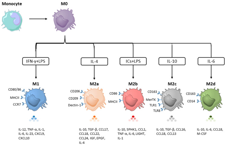
The tumor microenvironment (TME) is a critical regulator of tumor growth, progression, and metastasis. Among the innate immune cells recruited to the tumor site, macrophages are the most abundant cell population and are present at all stages of tumor progression. They undergo M1/M2 polarization in response to signals derived from TME. M1 macrophages suppress tumor growth, while their M2 counterparts exert pro-tumoral effects by promoting tumor growth, angiogenesis, metastasis, and resistance to current therapies. Several subsets of the M2 phenotype have been observed, often denoted as M2a, M2b, M2c, and M2d. These are induced by different stimuli and differ in phenotypes as well as functions. In this review, we discuss the key features of each M2 subset, their implications in cancers, and highlight the strategies that are being developed to harness TAMs for cancer treatment.
Keywords: cell signaling; polarization; solid cancers; targeted therapy; tumor microenvironment; tumor-associated macrophages.
Conflict of interest statement
The authors declare no conflict of interest.
Figures
Similar articles
-
Shaping Polarization Of Tumor-Associated Macrophages In Cancer Immunotherapy.Front Immunol. 2022 Jun 30;13:888713. doi: 10.3389/fimmu.2022.888713. eCollection 2022. Front Immunol. 2022. PMID: 35844605 Free PMC article. Review.
-
Dissection of pro-tumoral macrophage subtypes and immunosuppressive cells participating in M2 polarization.Inflamm Res. 2024 Sep;73(9):1411-1423. doi: 10.1007/s00011-024-01907-3. Epub 2024 Jun 27. Inflamm Res. 2024. PMID: 38935134 Free PMC article. Review.
-
Deciphering the performance of macrophages in tumour microenvironment: a call for precision immunotherapy.J Hematol Oncol. 2024 Jun 11;17(1):44. doi: 10.1186/s13045-024-01559-0. J Hematol Oncol. 2024. PMID: 38863020 Free PMC article. Review.
-
Macrophage Polarization States in the Tumor Microenvironment.Int J Mol Sci. 2021 Jun 29;22(13):6995. doi: 10.3390/ijms22136995. Int J Mol Sci. 2021. PMID: 34209703 Free PMC article. Review.
-
Role of cancer cell-derived exosomal glycoproteins in macrophage polarization.Mol Biol Rep. 2025 May 10;52(1):451. doi: 10.1007/s11033-025-10535-x. Mol Biol Rep. 2025. PMID: 40347313 Review.
Cited by
-
Evidence for a Pro-Inflammatory State of Macrophages from Non-Obese Type-2 Diabetic Goto-Kakizaki Rats.Int J Mol Sci. 2024 Sep 24;25(19):10240. doi: 10.3390/ijms251910240. Int J Mol Sci. 2024. PMID: 39408569 Free PMC article.
-
Gene Expression Analysis of (Paired) Primary and Relapsed Wilms Tumor Samples to Unravel the Underlying Factors Driving Tumor Recurrence.Cancer Med. 2025 Jun;14(11):e70969. doi: 10.1002/cam4.70969. Cancer Med. 2025. PMID: 40439002 Free PMC article.
-
The supporting role of Schwann cells in perineural invasion of pancreatic ductal adenocarcinoma.Front Pharmacol. 2025 Jun 11;16:1540027. doi: 10.3389/fphar.2025.1540027. eCollection 2025. Front Pharmacol. 2025. PMID: 40567365 Free PMC article. Review.
-
Emerging Advancements in Metabolic Properties of Macrophages within Disease Microenvironment for Immune Therapy.J Innate Immun. 2025;17(1):320-340. doi: 10.1159/000546476. Epub 2025 Jun 11. J Innate Immun. 2025. PMID: 40499523 Free PMC article. Review.
-
Identification and validation of M2 macrophage-related gene signature as a novel prognostic model for head and neck squamous cell carcinoma.Sci Rep. 2024 Oct 25;14(1):25338. doi: 10.1038/s41598-024-76866-0. Sci Rep. 2024. PMID: 39455885 Free PMC article.
References
-
- Liu Y., Ji X., Kang N., Zhou J., Liang X., Li J., Han T., Zhao C., Yang T. Tumor necrosis factor α inhibition overcomes immunosuppressive M2b macrophage-induced bevacizumab resistance in triple-negative breast cancer. Cell Death Dis. 2020;11:993. doi: 10.1038/s41419-020-03161-x. - DOI - PMC - PubMed
Publication types
MeSH terms
Grants and funding
LinkOut - more resources
Full Text Sources
Medical
