. 2023 Apr 19;24(8):7519.
doi: 10.3390/ijms24087519.

Effects of Nitric Oxide on the Activity of P2X and TRPV1 Receptors in Rat Meningeal Afferents of the Trigeminal Nerve

Affiliations

Effects of Nitric Oxide on the Activity of P2X and TRPV1 Receptors in Rat Meningeal Afferents of the Trigeminal Nerve

Kseniia Koroleva et al. Int J Mol Sci. .

Abstract

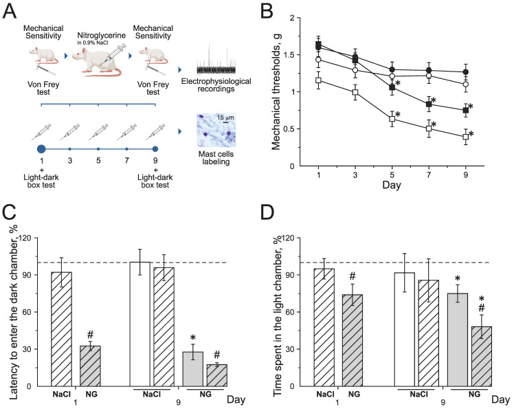
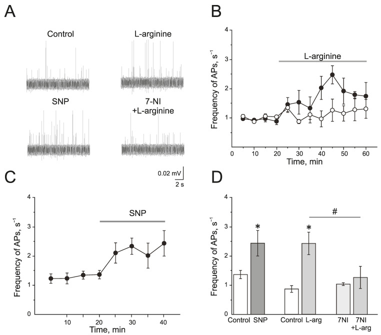
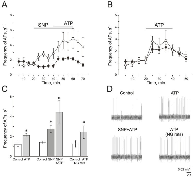
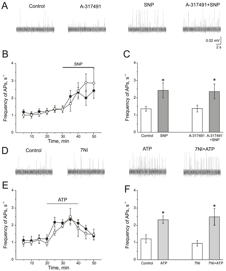
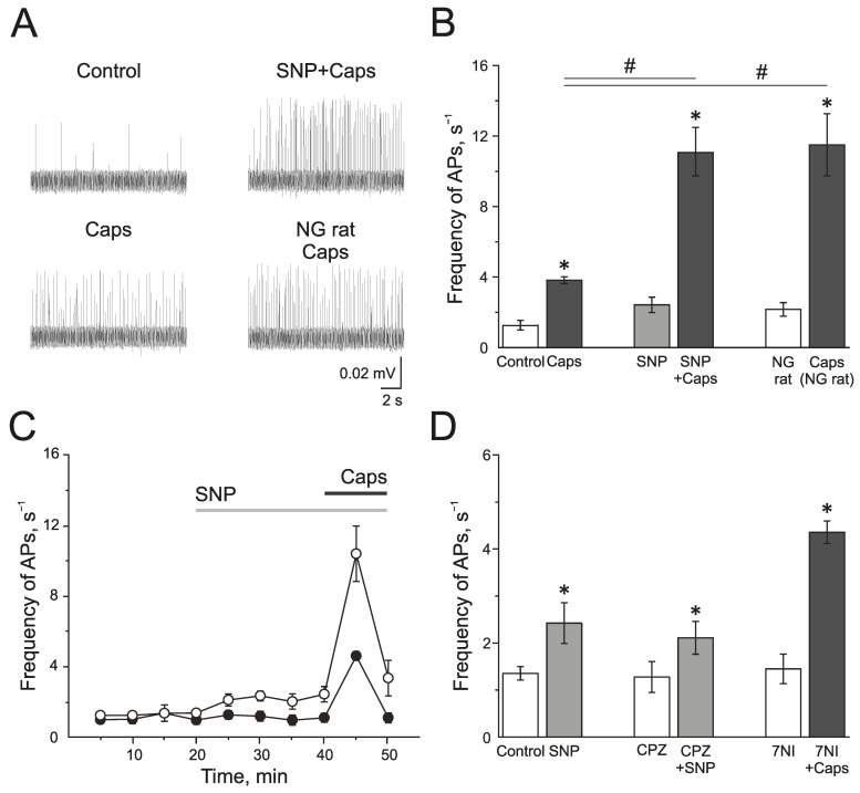
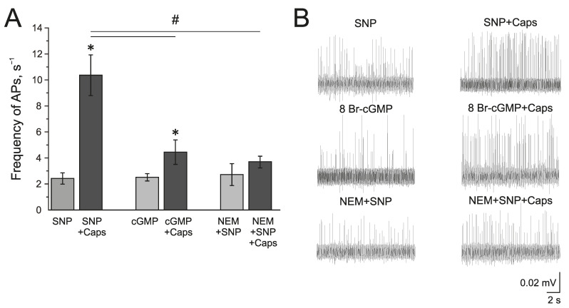
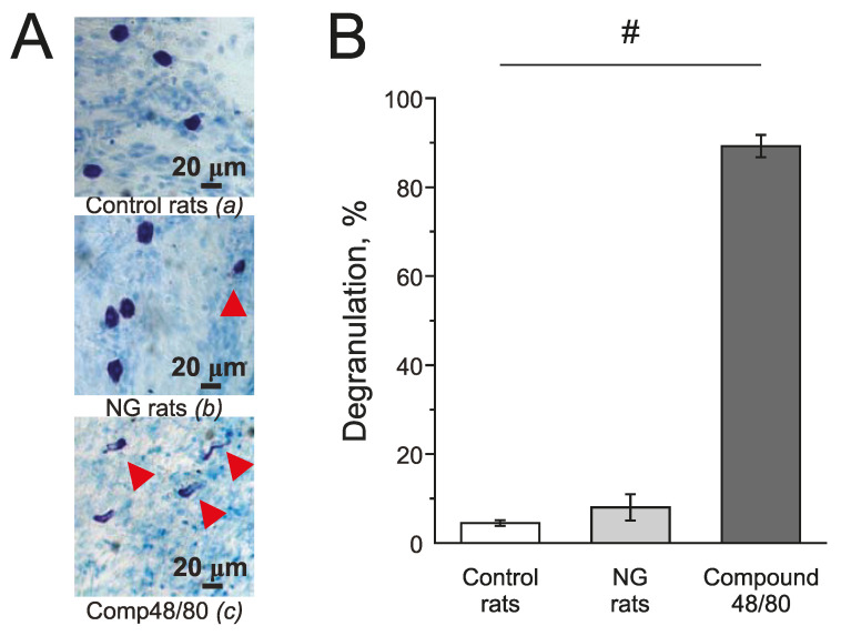
Nitric oxide is one of the endogenous molecules that play a key role in migraine. However, the interaction between NO and the main players in the nociceptive activity of the meningeal trigeminal afferents-TRPV1 and P2X3 receptors-remains unstudied. In the current project, the effects of acute and chronic NO administration on the activity of TRPV1 and P2X3 receptors in the peripheral afferents were studied using electrophysiological recording of action potentials of the trigeminal nerve in the rat hemiskull preparations. The data obtained indicate that exogenous and endogenous NO increased the activity of the trigeminal nerve independent on the inhibition of the TRPV1 and P2X3 receptors. The activity of the trigeminal nerve triggered by ATP changed neither in acute incubation in the NO donor-sodium nitroprusside (SNP) nor in the chronic nitroglycerine (NG)-induced migraine model. Moreover, the chronic NG administration did not increase in the number of degranulated mast cells in the rat meninges. At the same time, the capsaicin-induced activity of the trigeminal nerve was higher with chronic NO administration or after acute NO application, and these effects were prevented by N-ethylmaleimide. In conclusion, we suggested that NO positively modulates the activity of TRPV1 receptors by S-nitrosylation, which may contribute to the pro-nociceptive action of NO and underlie the sensitization of meningeal afferents in chronic migraine.

Keywords: P2X3 receptor; S-nitrosylation; TRPV1; mast cell; migraine; nitric oxide; nitroglycerine; trigeminal nerve.


Conflict of interest statement

The authors declare no conflict of interest.

Figures

Figure 1
Figure 1
Effect of chronic nitroglycerin (NG) administration on mechanical sensitivity and photophobia. (A) Experimental scheme. (B) Mechanical withdrawal thresholds of rats before and two hours after the administration of NG (black and white squares) or vehicle (NaCl) (black and white circles). (C) Latency to enter the dark chamber and (D) time spent in the light chamber before (open columns) and two hours after (dashed columns) the administration of vehicle (white columns) or NG (grey columns) on the 1st and 9th day (in %) compared to the initial values taken as 100% and marked with a dotted line. * p < 0.05 compared to 100%; # p < 0.05 compared to the pre-injection level.
Figure 2
Figure 2
Endogenous and exogenous nitric oxide (NO) increases the electrical activity of rat trigeminal afferents. (A) Sample traces of AP in the trigeminal nerve of a rat in the control and after the application of L-arginine (300 μM), NO donor SNP (200 μM), and L-arginine in the presence of nNOS inhibitor, 7-nitroindazole (7NI, 1 mM); (B) The time-course of AP frequency after the application of L-arginine (black circles) and in the presence of 7NI (white circles); (C) The time-course of frequency of AP after the application of SNP; (D) Histogram showing the maximum frequency of AP per 5 min after the application of L-arginine, SNP, 7NI and 7NI + L-arginine. Mean ± SEM. * p < 0.05 compared to control values; # p < 0.05 compared to the effect of L-arginine as a control.
Figure 3
Figure 3
The role of NO in the pro-nociceptive action of ATP in the trigeminal nerve afferents. (A) The time-course of AP frequency during ATP (100 µM) application in the control group (black circles) and in the group after the application of the NO donor (SNP 200 µM, white circles); (B) The time-course of the AP frequency during the application of ATP in the control (black circles) and in the nitroglycerine (NG)-induced migraine model (white circles); (C) Histogram showing the maximum frequency of AP per 5 min in the control, after the application of ATP; SNP, ATP in the presence of SNP and the effect of ATP in the NG-induced migraine model group (NG rats); (D) Sample traces of AP in the trigeminal nerve in the control, after the application of ATP, application of ATP in the presence of SNP, and application of ATP in the NG-induced migraine model group (NG rats). Mean ± SEM. * p < 0.05.
Figure 4
Figure 4
Interaction of NO and P2X3 receptors in rat trigeminal afferents (A) Sample traces of AP in the trigeminal nerve in the control or after the inhibition of P2X3 receptors by A-317491 (10 µM), after the application of the NO donor—SNP (200 µM) and A-317491 + SNP; (B) The time-course of AP frequency after the application of SNP in control (black circles) and after the inhibition of P2X3 receptors by A-317491 (white circles). (C) Histogram showing the maximum frequency of AP per 5 min in the trigeminal nerve after the application of SNP and A-31749 + SNP. (D) Sample traces of AP in the control, after the application of 7NI, ATP and 7NI + ATP; (E) The time-course of AP frequency after the application of ATP in the control (black circles) and in the presence of 7NI (white circles); (F) Histogram showing the maximum frequency of AP per 5 min after the application of ATP in the control and in the presence of 7NI. Mean ± SEM. * p < 0.05.
Figure 5
Figure 5
Effects of NO on activity of TRPV1 in the rat trigeminal afferents. (A) Sample traces of AP in the trigeminal nerve in controls, after the application of capsaicin (1 µM), capsaicin in the presence of NO donor, SNP (200 µM), and the application of capsaicin in NG-induced migraine model group. (B) Histogram showing the mean frequency of AP per 5 min in the trigeminal nerve after the application of capsaicin in the control, after application of the NO donor SNP and in the NG-induced migraine model group; (C) The time-course of frequency of AP after the application of capsaicin in the control (black circles) and after the application of SNP (white circles); (D) Histogram showing the mean frequency of AP per 5 min after the application of SNP in the control group, application of SNP after the inhibition of TRPV1 receptors by capsazepine (20 µM) and capsaicin after inhibition of nNOS by 7NI (1 mM). Mean ± SEM. * p < 0.05 (compared to the control); # p < 0.05 (compared to the effect of capsaicin in the control).
Figure 6
Figure 6
Mechanism of the modulating effect of NO on TRPV1 receptors. (A) Histogram showing the maximum frequency of AP per 5 min in the trigeminal nerve after the application of the NO donor SNP (200 μM) and capsaicin (1 μM) in the presence of SNP; cGMP analogue—8-Br-cGMP (500 μM) and capsaicin in the presence of 8-Br-cGMP; N-Ethylmaleimide, (NEM, 25 μM) together with SNP and capsaicin in the presence of NEM + SNP. (B) Sample traces of AP in the trigeminal nerve after the application of SNP and SNP + Caps; 8-Br-cGMP and 8-Br-cGMP + Caps; NEM and NEM + SNP + Caps. Mean ± SEM. * p < 0.05 (compared to SNP or cGMP respectively); # p < 0.05 (compared to the effect of capsaicin + SNP).
Figure 7
Figure 7
Effect of chronic nitroglycerine administration on mast cell degranulation in rat meninges. (A) Toluidine Blue staining of the meninges in the control (a) (n = 5); in the NG-induced migraine model group (NG rats; n = 4); (b); after exposure to Compound 48/80 in the control (10 mg/mL; 30 min; n = 4) (c). Notice red arrows indicating degranulated mast cells. (B) Histograms showing the percent of degranulated mast cells in the control group, model group, and after Compound 48/80. Mean ± SEM. # p < 0.05.

Similar articles

Cited by

References

    1. Hermann A., Sitdikova G.F., Weiger T.M. Gasotransmitters: Physiology and Pathophysiology. Springer; Berlin, Heidelberg, Germany: 2012. pp. 1–204. - DOI
    1. Picón-Pagès P., Garcia-Buendia J., Muñoz F.J. Functions and Dysfunctions of Nitric Oxide in Brain. Biochim. Biophys. Acta-Mol. Basis Dis. 2019;1865:1949–1967. doi: 10.1016/j.bbadis.2018.11.007. - DOI - PubMed
    1. Cyr A.R., Huckaby L.V., Shiva S.S., Zuckerbraun B.S. Nitric Oxide and Endothelial Dysfunction. Crit. Care Clin. 2020;36:307–321. doi: 10.1016/j.ccc.2019.12.009. - DOI - PMC - PubMed
    1. Lundberg J.O., Weitzberg E. Nitric Oxide Signaling in Health and Disease. Cell. 2022;185:2853–2878. doi: 10.1016/j.cell.2022.06.010. - DOI - PubMed
    1. Ghimire K., Altmann H.M., Straub A.C., Isenberg J.S. Nitric Oxide: What’s New to NO? Am. J. Physiol.-Cell Physiol. 2017;312:C254–C262. doi: 10.1152/ajpcell.00315.2016. - DOI - PMC - PubMed