. 2023 Apr 19;24(8):7525.
doi: 10.3390/ijms24087525.

Multiple Nucleocapsid Structural Forms of Shrimp White Spot Syndrome Virus Suggests a Novel Viral Morphogenetic Pathway

Affiliations

Multiple Nucleocapsid Structural Forms of Shrimp White Spot Syndrome Virus Suggests a Novel Viral Morphogenetic Pathway

Hui-Ju Huang et al. Int J Mol Sci. .

Abstract

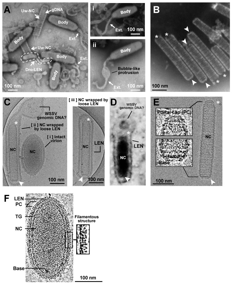
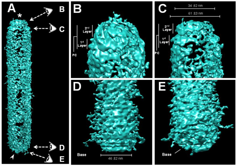
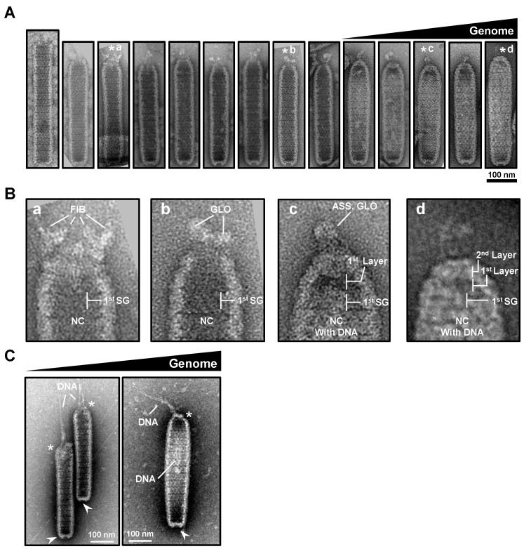
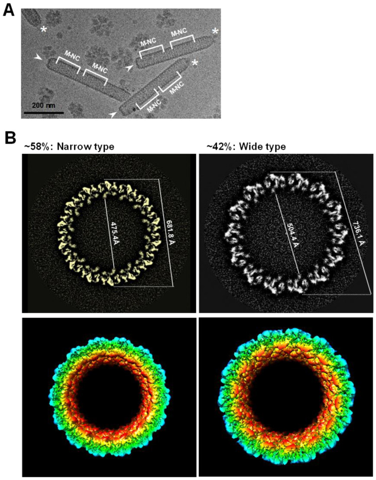
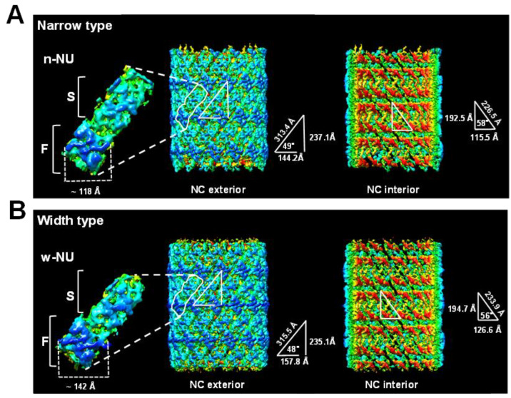
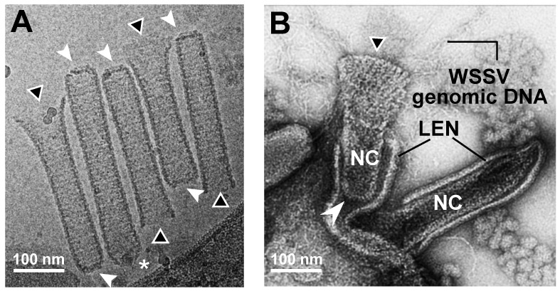
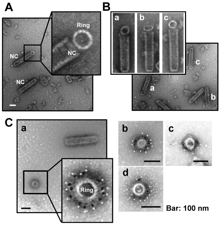
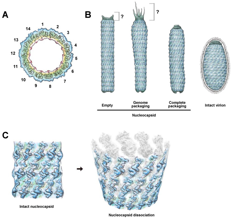
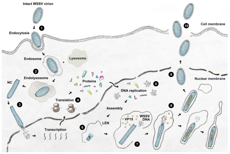
White spot syndrome virus (WSSV) is a very large dsDNA virus. The accepted shape of the WSSV virion has been as ellipsoidal, with a tail-like extension. However, due to the scarcity of reliable references, the pathogenesis and morphogenesis of WSSV are not well understood. Here, we used transmission electron microscopy (TEM) and cryogenic electron microscopy (Cryo-EM) to address some knowledge gaps. We concluded that mature WSSV virions with a stout oval-like shape do not have tail-like extensions. Furthermore, there were two distinct ends in WSSV nucleocapsids: a portal cap and a closed base. A C14 symmetric structure of the WSSV nucleocapsid was also proposed, according to our Cryo-EM map. Immunoelectron microscopy (IEM) revealed that VP664 proteins, the main components of the 14 assembly units, form a ring-like architecture. Moreover, WSSV nucleocapsids were also observed to undergo unique helical dissociation. Based on these new results, we propose a novel morphogenetic pathway of WSSV.

Keywords: cryogenic electron microscopy; large dsDNA virus; nucleocapsid; white spot syndrome virus.


Conflict of interest statement

The authors declare no conflict of interest.

Figures

Figure 1
Figure 1
Images of WSSV enveloped virion and nucleocapsid captured with PTA-negative staining TEM and Cryo-EM. (A) With TEM, WSSV virions with a long oval-shaped body (Body) and with/without a flexible tail-like extension (Ext.) were observed. The dotted-line area indicates a dropped lipid-containing envelope (Dro-LEN). The nucleocapsid unwrapped by LEN (Uw-NC) indicates the prominence where the viral DNA genome (gDNAs) is aggregated. There were two types of WSSV virion with a tail-like extension (Ai,Aii). (B) TEM images of purified nucleocapsid without viral DNA genome (Empty NC) show a striped tube-like structure with two ends: an open portal cap end (* asterisk) and a closed basal end (arrow). (C) With Cryo-EM, three types of enveloped WSSV virions with the nucleocapsid (NC) were observed: [i] a shortened, oval-shaped virion with an oval-shaped NC wrapped by a firm envelope; [ii] a long virion with a striped tube NC, wrapped by a loose extra-extended envelope and an unknown aggregation (may be a WSSV genomic DNA with viral DNA-binding proteins); and [iii] a long virion with a striped tube NC wrapped by a loose envelope. The nucleocapsids had two distinct ends: the portal caps and the closed-base ends, denoted by asterisks and arrows, respectively. (D) TEM image of WSSV virion similar to Figure 1Cii in the nucleus of WSSV-infected cells. (E) With Cryo-EM, the purified striped tube-like nucleocapsid clearly had a portal cap structure (*) and closed basal end (arrow). (F) Cryo-EM image of the intact WSSV virion particle. The intact virion has three layers: a lipid-containing envelope (LEN), tegument (TG), and nucleocapsid (NC). The portal cap structure (PC) and closed basal end (Base) of the nucleocapsid were also observed. Filamentous structures on the surface of the lipid-containing envelope (LEN).
Figure 2
Figure 2
Three-dimensional volume reconstruction of WSSV nucleocapsid by cryo-electron tomography (Cryo-ET). (A) The reconstructed Cryo-ET of the WSSV nucleocapsid with a portal cap structure (*) and a closed basal end (arrow); the four eyes represent distinct visual angles shown in Figure (BE); (B,C) the portal cap structure with two layers; (D,E) the closed basal end had a concave plate shape.
Figure 3
Figure 3
A proposed model of the WSSV nucleocapsid revealing its DNA packaging machinery based on TEM images. (A) Purified nucleocapsids negative-stained with UA or UF in the TEM micrographs revealed a possible process of viral DNA packaging from the opening portal cap region to form a mature intact nucleocapsid with WSSV genomic DNAs (the mist in the nucleocapsid). Potential contraction and capped events to close the opening portal region were also proposed for the nucleocapsid to assemble properly. The images were organized arbitrarily based on the cap formation, shape of the nucleocapsid, and amount of DNA packaged in the nucleocapsid. The cap ends of the nucleocapsids (labeled with asterisks) were selected in (B); (B) capped events of the opening portal region of the nucleocapsids (NC). The sequential portal cap formation is shown in B (ad). First SG: the first segment of the nucleocapsid (NC); FIB: fiber-like structures; GLO: globin-like structure; ASS. GLO: assembled globin-like structure. The 1st and 2nd layers of the portal cap were designated based on Figure 2. (C) The potential event of WSSV DNA translocation through the portal cap end of the nucleocapsid.
Figure 4
Figure 4
A near C14-symmetry of WSSV nucleocapsid. (A) Selected regions suitable for high-resolution Cryo-EM structure of WSSV nucleocapsids. After purifying WSSV nucleocapsids, the middle regions of WSSV nucleocapsid (M-NC) with relatively uniform structures were subjected to the construction of tomographic images; (B) diagnostic plot detail to interpret top views of the middle part of WSSV nucleocapsids (M-NC) in two packing types. Based on all the WSSV nucleocapsids evaluated, ~58% were the narrow type, whereas ~42% were the wide type. The regular feature, i.e., ‘the C14-symmetry’ of the WSSV nucleocapsid, was observed in both types. The corresponding external diameters and internal diameters of each NC type were measured.
Figure 5
Figure 5
Exterior and interior surfaces of WSSV nucleocapsid (A) narrow type and (B) wide type. The left part of the images is the outer nucleocapsid surface, whereas the right part is the inner side. All the structures were radially colored from the origin (red) to the outer surface (deep blue). Nucleocapsid units (NU) with flower (F) and stem (S) domains from each type are also shown. These images were generated using CHIMERA software version 1.13.1.
Figure 6
Figure 6
Images of dissociation of WSSV nucleocapsids. (A) Dissociation of WSSV nucleocapsids was initiated at the portal cap end. This image was captured with Cryo-EM; (B) dissociation of WSSV nucleocapsids as 14-helical strands. This image was captured with UA-negative staining TEM. *: portal cap structure; ▲: dissociated cap end; arrow: closed base end.
Figure 7
Figure 7
Immunoelectron microscopy analysis of purified nucleocapsid probed with a VP664 antibody. (A) Purified WSSV nucleocapsids had a ring-like structure. (B) Ring-like structures were observed in the portal cap end. (C) Most gold particles (asterisks) were specifically localized to the exterior side of these ring-like structures.
Figure 8
Figure 8
Schematic illustrations of (A) overall C14 symmetry of WSSV nucleocapsid; (B) process from empty nucleocapsids, through nucleocapsids with WSSV genomic DNAs, to intact virion with lipid-containing envelope; and (C) a proposed 14-helical-strand dissociation model of WSSV nucleocapsids. Note: these drawings are hand-drawn and not computer-generated.
Figure 9
Figure 9
A proposed model of WSSV entry, maturation, and morphogenesis, with the following steps: (1) WSSV entry into the cell; (2) stepwise disassembly process from intact virion to nucleocapsid in endosome-lysosome fusion; (3) naked nucleocapsids presumably undergo the release of WSSV genomes from the portal cap end into the nucleus; (4) transcription and translation of WSSV genes begin when the virogenic stroma is formed; (5) replication of WSSV genomes; (6) structural proteins of envelope, tegument, and nucleocapsids start to assemble; (7) WSSV genomes with some viral DNA-binding proteins are packaged into a mature capsid through the portal cap; (8) envelope precursors begin to shrink and remove excess parts to fit the nucleocapsid; (9) intact WSSV virions are completely assembled in the nucleus; (10) intact WSSV virions are released after cell lysis.

Similar articles

Cited by

References

    1. Dey B.K., Dugassa G.H., Hinzano S.M., Bossier P. Causative agent, diagnosis and management of white spot disease in shrimp: A review. Rev. Aquac. 2020;12:822–865. doi: 10.1111/raq.12352. - DOI
    1. Chou H.Y., Huang C.Y., Wang C.H., Chiang H.C., Lo C.F. Pathogenicity of a baculovirus infection causing White Spot Syndrome in cultured Penaeid shrimp in Taiwan. Dis. Aquat. Organ. 1995;23:165–173. doi: 10.3354/dao023165. - DOI
    1. Yang F., He J., Lin X., Li Q., Pan D., Zhang X., Xu X. Complete genome sequence of shrimp white spot bacilliform virus. J. Virol. 2001;75:11811–11820. doi: 10.1128/JVI.75.23.11811-11820.2001. - DOI - PMC - PubMed
    1. Chen L.L., Wang H.C., Huang C.J., Peng S.E., Chen Y.G., Lin S.J., Chen W.Y., Dai C.F., Yu H.T., Wang C.H., et al. Transcriptional analysis of the DNA polymerase gene of shrimp white spot syndrome virus (WSSV) Virology. 2002;301:136–147. doi: 10.1006/viro.2002.1536. - DOI - PubMed
    1. Wang H.C., Hirono I., Maningas M.B.B., Somboonwiwat K., Stentiford G. ICTV Report Consortium. ICTV Virus Taxonomy Profile: Nimaviridae. J. Gen. Virol. 2019;100:1053–1054. doi: 10.1099/jgv.0.001248. - DOI - PubMed

LinkOut - more resources