. 2023 Apr 19;24(8):7528.
doi: 10.3390/ijms24087528.

JI017 Induces Cell Autophagy and Apoptosis via Elevated Levels of Reactive Oxygen Species in Human Lung Cancer Cells

Affiliations

JI017 Induces Cell Autophagy and Apoptosis via Elevated Levels of Reactive Oxygen Species in Human Lung Cancer Cells

Jin Mo Ku et al. Int J Mol Sci. .

Abstract

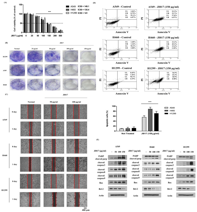
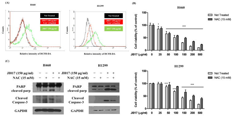
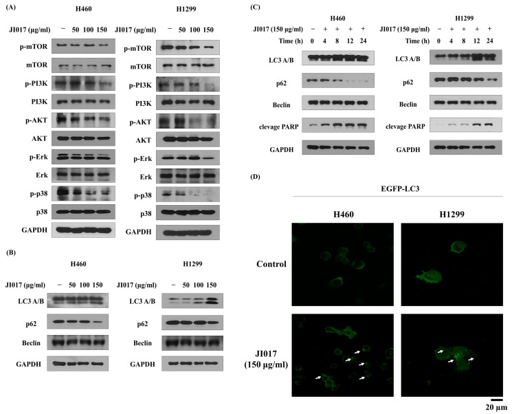
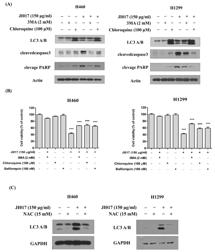
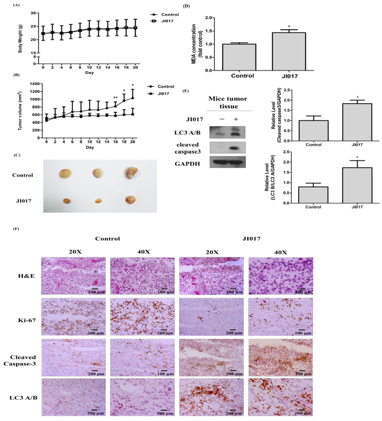
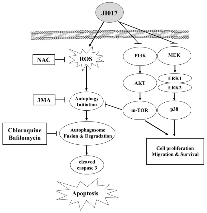
Lung cancer is one of the most common malignant tumors and a leading cause of cancer-related death in the worldwide. Various anticancer drugs, such as cisplatin and pemetrexed, have been developed for lung cancer treatment but due their drug resistance and side effects, novel treatments need to be developed. In this study, the efficacy of the natural drug JI017, which is known to have few side effects, was tested in lung cancer cells. JI017 inhibited A549, H460, and H1299 cell proliferation. JI017 induced apoptosis, regulated apoptotic molecules, and inhibited colony formation. Additionally, JI017 increased intracellular ROS generation. JI017 downregulated PI3K, AKT, and mTOR expression. JI017 increased the cytosolic accumulation of LC3. We found that JI017 promoted apoptosis through ROS-induced autophagy. Additionally, the xenograft tumor size was smaller in JI017-treated mice. We found that JI017 treatment increased MDA concentrations, decreased Ki-67 protein levels, and increased cleaved caspase-3 and LC3 levels in vivo. JI017 decreased cell proliferation and increased apoptosis by inducing autophagy signaling in H460 and H1299 lung cancer cells. Targeting JI017 and autophagy signaling could be useful in lung cancer treatment.

Keywords: JI017; ROS; autophagy; lung cancer.


Conflict of interest statement

The authors declare no conflict of interest.

Figures

Figure 1
Figure 1
Effect of JI017 on A549, H460, and H1299 cell viability. (A) Cells were treated with different concentrations of JI017 for 24 h. (B) Cell viability was then measured using the MTS assay. H460, A549, and H1299 cells were treated with JI017 and exposed for 7 days, then the effect on cell growth was assessed using a colony formation assay. (C) The migration of JI017-treated cells was assessed using a wound-healing assay. Intervals between cells are marked with red lines. Scale bar = 400 µm. (D) H460 and H1299 cells were treated with JI017 for 24 h, stained with Annexin V/PI, and analyzed by flow cytometry. (E) Whole cell lysates were analyzed by Western blotting with anti-PARP; anti-cleaved caspase 3, -8, and -9; and anti-Bax, anti-Bcl-2, and anti-Actin antibodies. Data are presented as the mean ± SEM. *** p < 0.001 compared to untreated cells.
Figure 2
Figure 2
Effect of JI017 in A549, H460, and H1299 ROS accumulation. (A) Treatment with JI017 (150 μg/mL) in H460 and H1299 cells for 24 h; they were labeled with DCFH-DA (10 μM) for 30 min. Additionally, intracellular ROS levels were determined by flow cytometry. Pretreatment with NAC (15 mM) for 1 h was followed by treatment with various concentrations of JI017. (B) Cell viability was measured using the MTS assay. (C) Whole cell lysates were analyzed by Western blotting with anti-PARP, anti-cleaved caspase 3, and anti-GAPDH antibodies. Data are presented as the mean ± SEM. * p < 0.05 and *** p < 0.001 compared to untreated cells.
Figure 3
Figure 3
JI017 activated autophagy by suppressing the PI3K-AKT-mTOR pathway in H460 and H1299 cells. H460 and H1299 cells were treated with JI017 (50, 100, and 150 μg/mL) for 24 h. (A,B) Whole cell lysates were analyzed by Western blotting. (C) H460 and H1299 cells were treated with JI017 (150 ng/mL) for different times (4, 8, 12, and 24 h). (D) H460 and H1299 cells transfected with the pEGFP-LC3 vector were treated with JI017 (150 μg/mL) for 8 h. The Fluorescence microscopy analysis confirmed positive staining of LC3B puncta. LC3B puncta are indicated by white arrows. Scale bar = 20 µm.
Figure 4
Figure 4
JI017 induced apoptosis through an autophagy pathway and ROS generation. (A) H460 and H1299 cells were treated with 3MA (2 mM) or chloroquine (100 μM) combined with JI017 (150 μg/mL). 3MA, chloroquine, or bafilomycin treatment was followed by treatment with JI017. (B) Cell viability was measured using MTS assay. (C) H460 and H1299 cells were treated with NAC (15 mM) and JI017 (150 μg/mL). Whole cell lysates were analyzed by Western blotting with anti-LC3 and anti-GAPDH antibodies. Data are presented as the mean ± SEM. *** p < 0.001 compared to untreated cells.
Figure 5
Figure 5
JI017 suppressed lung cancer cell growth in mice. BALB/c nude mice were subcutaneously injected with H460 cells. (A) The mouse body weight and (B) the tumor growth rate are shown. (C) Representative tumor images of the control group and the JI017-treated group. (D) The MDA concentration was assessed by a thiobarbituric reactive substance (TBARS) assay and normalized to the protein concentration. (E) Whole tissue lysates were analyzed by Western blotting with anti-LC3, anti-cleaved caspase 3, and anti-GAPDH antibodies. (F) The IHC staining of Ki-67 and cleaved caspase-3 and LC3 was carried out. Scale bar = 200 µm for 20× and Scale bar = 100 µm for 40×. The data are expressed as the mean ± SEM in all groups (n = 8–11). * p < 0.05 and ** p < 0.01 compared to the untreated group.
Figure 6
Figure 6
Mechanistic pathway diagram for the autophagy activation potency of JI017 through AKT, ERK inhibition, and ROS induction in lung cancer.

References

    1. Goodman J.E., Mayfield D.B., Becker R.A., Hartigan S.B., Erraguntla N.K. Recommendations for further revisions to improve the International Agency for Research on Cancer (IARC) Monograph program. Regul. Toxicol. Pharmacol. 2020;113:104639. doi: 10.1016/j.yrtph.2020.104639. - DOI - PubMed
    1. Griffin R., Ramirez R.A. Molecular Targets in Non-Small Cell Lung Cancer. Ochsner J. 2017;17:388–392. - PMC - PubMed
    1. Miller H.A., van Berkel V.H., Frieboes H.B. Lung cancer survival prediction and biomarker identification with an ensemble machine learning analysis of tumor core biopsy metabolomic data. Metab. Off. J. Metab. Soc. 2022;18:57. doi: 10.1007/s11306-022-01918-3. - DOI - PMC - PubMed
    1. Iqbal N., Shukla N.K., Deo S.V., Agarwala S., Sharma D.N., Sharma M.C., Bakhshi S. Prognostic factors affecting survival in metastatic soft tissue sarcoma: An analysis of 110 patients. Clin. Transl. Oncol. 2016;18:310–316. doi: 10.1007/s12094-015-1369-9. - DOI - PubMed
    1. Jonna S., Subramaniam D.S. Molecular diagnostics and targeted therapies in non-small cell lung cancer (NSCLC): An update. Discov. Med. 2019;27:167–170. - PubMed

Substances