. 2023 Apr 19;24(8):7543.
doi: 10.3390/ijms24087543.

Characterization of Porcine Ocular Surface Epithelial Microenvironment

Affiliations

Characterization of Porcine Ocular Surface Epithelial Microenvironment

Naresh Polisetti et al. Int J Mol Sci. .

Abstract

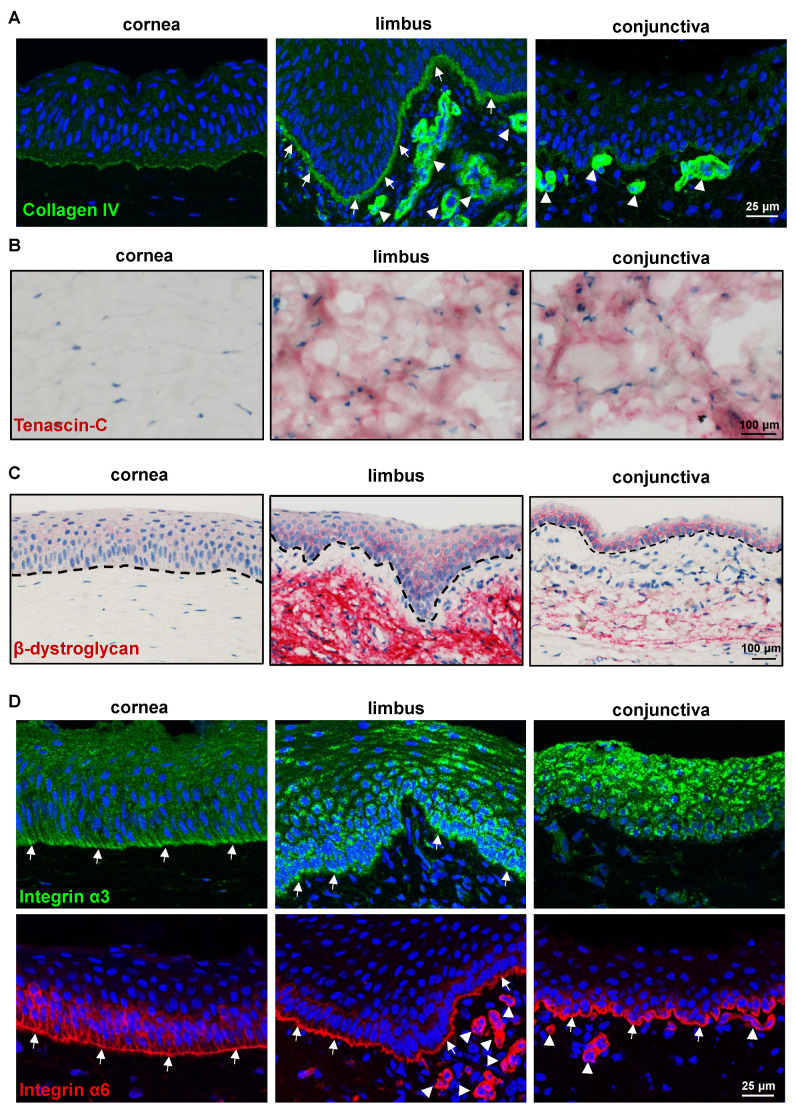
The porcine ocular surface is used as a model of the human ocular surface; however, a detailed characterization of the porcine ocular surface has not been documented. This is due, in part, to the scarcity of antibodies produced specifically against the porcine ocular surface cell types or structures. We performed a histological and immunohistochemical investigation on frozen and formalin-fixed, paraffin-embedded ocular surface tissue from domestic pigs using a panel of 41 different antibodies related to epithelial progenitor/differentiation phenotypes, extracellular matrix and associated molecules, and various niche cell types. Our observations suggested that the Bowman's layer is not evident in the cornea; the deep invaginations of the limbal epithelium in the limbal zone are analogous to the limbal interpalisade crypts of human limbal tissue; and the presence of goblet cells in the bulbar conjunctiva. Immunohistochemistry analysis revealed that the epithelial progenitor markers cytokeratin (CK)15, CK14, p63α, and P-cadherin were expressed in both the limbal and conjunctival basal epithelium, whereas the basal cells of the limbal and conjunctival epithelium did not stain for CK3, CK12, E-cadherin, and CK13. Antibodies detecting marker proteins related to the extracellular matrix (collagen IV, Tenascin-C), cell-matrix adhesion (β-dystroglycan, integrin α3 and α6), mesenchymal cells (vimentin, CD90, CD44), neurons (neurofilament), immune cells (HLA-ABC; HLA-DR, CD1, CD4, CD14), vasculature (von Willebrand factor), and melanocytes (SRY-homeobox-10, human melanoma black-45, Tyrosinase) on the normal human ocular surface demonstrated similar immunoreactivity on the normal porcine ocular surface. Only a few antibodies (directed against N-cadherin, fibronectin, agrin, laminin α3 and α5, melan-A) appeared unreactive on porcine tissues. Our findings characterize the main immunohistochemical properties of the porcine ocular surface and provide a morphological and immunohistochemical basis useful to research using porcine models. Furthermore, the analyzed porcine ocular structures are similar to those of humans, confirming the potential usefulness of pig eyes to study ocular surface physiology and pathophysiology.

Keywords: conjunctiva; cornea; extracellular matrix; limbal epithelial progenitor cells; limbal niche cells; limbal stem cells; melanocytes; porcine ocular surface.


Conflict of interest statement

The authors declare no conflict of interest.

Figures

Figure 1
Figure 1
Histology of porcine ocular surface: (A) Light micrographs of paraffin sections of corneoconjunctival tissue stained with hematoxylin and eosin (HE) showing the corneal epithelium consists of 6–8 layers of stratified epithelium and Bowman’s layer is not evident; deep invaginations of the limbal epithelium (6–12 layers) analogous to the limbal interpalisade crypts of human limbal tissue can be observed in the limbal zone; the bulbar conjunctiva is composed of a 3–5 layered stratified epithelium; significant vessels (arrows) in the limbal and conjunctival stroma. The conjunctival and limbal stroma are organized differently to the corneal stroma, with loose connective tissue, a large number of fibroblasts, and the significant presence of vessels (arrows). Alcian blue (AB) staining of ocular surface showing the presence of glycosaminoglycans (blue) throughout the cornea and rather weak staining in the limbal and conjunctival stroma and the presence of goblet cells in the conjunctival epithelium (arrows). Periodic acid-Schiff (PAS) staining highlighting the BM of porcine corneal epithelium (arrows) and less pronounced in the epithelial BM of limbus and conjunctiva. (B) Transmission electron micrographs showing the architecture of the corneal (i), limbal (ii), and the presence of goblet cells in the conjunctival epithelia (arrows, (iii)) (dotted line represents basement membrane); extensive basal cell processes of limbal epithelial progenitor cells (LEPC) extending into the limbal stroma (ST) (arrows, (iv)), close interactions with stromal immune cells (arrows, (v)) and with mesenchymal stromal cells (arrows, (vi)).
Figure 2
Figure 2
Immunohistochemical analysis of epithelial-related markers: Immunohistochemical staining of paraffin sections showing the expression of epithelial keratins (pan-CK) in all epithelial layers of the cornea, limbus, and conjunctiva (red); PAX6 expression in all layers of corneal epithelium and in nearly all epithelial cell layers of limbus and conjunctiva except for some basal cells (white arrows) and superficial epithelial cells (black arrows); cytokeratin (CK) 12 expression in all layers of the corneal epithelium, but only in suprabasal cell layers of limbal epithelium (arrows); CK13 expression in the suprabasal cell layers of both limbal and conjunctival epithelium and negative in the basal layers (arrows); CK14 expression in the basal layers of the limbal and conjunctival epithelia (arrows) but negative in the corneal epithelium; and CK17/19 expression in basal layers of the limbal epithelium (arrows) and in all layers of the conjunctival epithelium (arrows); dashed line represents basement membrane.
Figure 3
Figure 3
Expression analysis of epithelial-related markers: (A) Immunostaining of corneoconjunctival tissue showing the expression of p63α in the basal layers of corneal, limbal, and conjunctival epithelia, but the stronger expression in the limbus and conjunctiva and a few negative cells in the limbal and conjunctival basal layers (arrows); Ki-67 expression in the basal cells of the cornea, limbus, and conjunctiva (arrows); dashed line represents basement membrane (BM). (B) Immunofluorescence staining of frozen sections showing the expression of E-cadherin in the superficial layers of epithelial cells in both limbus and conjunctiva (red), leaving the basal cells negative (arrows), whereas all layers of corneal epithelium expressed E-cadherin; CK15 expression in the basal layers of the limbal and conjunctival epithelia (arrows, red) but not in the corneal epithelia; P-cadherin expression in the basal layers of limbal and conjunctival epithelia (arrows, green); CK3/76 expression (green) in all layers of the corneal epithelium and in suprabasal cell layers of limbal epithelium but not in basal layers (arrows); nuclear counterstaining with 4′,6-diamidino-2-phenylindole (blue); dashed line represents BM.
Figure 4
Figure 4
Expression analysis of extracellular matrix and adhesion molecules: (A) Immunohistochemistry and laser confocal imaging of corneoconjunctival tissue sections showing the collagen IV strongly stained limbal epithelial basement membrane (BM) (arrows); very weak staining in corneal and conjunctival epithelial BM; strong staining in the limbal and conjunctival vessel BM (arrowheads). Nuclei are counterstained with 4′,6-diamidino-2-phenylindole (DAPI, blue). (B) Immunohistochemical analysis of corneoconjunctival tissue showing the tenascin(TN)-C expression in the stroma of limbus and conjunctiva (red) but not in the corneal stroma. (C) Immunohistochemical staining of corneoconjunctival tissue sections showing the β-dystroglycan expression in the basal cell membranes of the limbus and conjunctiva; faint membrane staining in the superficial cells of the limbus and conjunctiva as well as corneal epithelium; prominent staining in the limbal stroma but weak in the conjunctival stroma and absent in the corneal stroma. The dashed line represents BM. (D) Immunohistochemistry and laser confocal imaging of corneoconjunctival tissue sections showing the integrin α3 expression in the basal aspect of corneal, limbal, and conjunctival epithelium, with the limbus and conjunctiva showing a strong expression (green); integrin α6 expression in all ocular surface epithelia, with a continuous strong basal membrane staining (arrows) and prominent staining in the vasculature of limbal and conjunctival stroma (arrowheads); nuclei are counterstained with DAPI (blue).
Figure 5
Figure 5
Expression analysis of limbal niche cell-related markers: (A) Immunohistochemical staining of paraffin sections of corneoconjunctival specimen showing the vimentin expression in the stromal cells of the porcine cornea, limbus, and conjunctiva (red); vimentin+ cells in the basal epithelial layers of both limbus and conjunctiva (arrowheads); dashed line represents the basement membrane (BM). (B) Immunofluorescence staining corneoconjunctival tissue sections showing the CD44 expression on the plasma membranes of basal epithelial and stromal cells of the cornea (red) and predominant expression in the limbus and conjunctiva; CD90 expression in the stromal cells of limbus and conjunctiva (red); neurofilament (NF) expression in the stroma of all three surface regions (arrowheads) and in the limbal BM (arrowheads); the anti-human HLA-class I antibody stained all cells of the ocular surface (dashed line represents BM).
Figure 6
Figure 6
Expression analysis of immune-related markers: (A) Immunohistochemical staining of porcine ocular surface paraffin tissue sections showing the swine leukocyte class II+ cells (detected by HLA-class II antibody) in the epithelial layers and stroma of limbus and conjunctiva (arrows); S100 expression in a few cells of the limbus and the conjunctival epithelial basal layer (arrowheads) and in the stroma of all three regions (arrows); anti-von Willebrand factor (VWF) stained the vasculature of the porcine ocular limbus and conjunctiva (arrows); expression CD4 in a few cells of limbal and conjunctival stroma; dashed line represent basement membrane (BM). (B) Immunofluorescence staining of frozen sections showing the CD1+ cells within the multilayered epithelium of limbus and conjunctiva (arrows) but not in the cornea; CD14 expression in a few cells of the limbal and conjunctival stroma (arrows), rarely observed only in the conjunctival epithelium (arrow); nuclei are counterstained with 4′,6-diamidino-2-phenylindole (blue); dashed line represents the BM.
Figure 7
Figure 7
Expression analysis of melanocyte-related markers: (A) Light micrographs of paraffin sections of corneoconjunctival specimen stained with Fontana-Masson showing the melanin in the limbal and conjunctival regions of domestic porcine samples and wild pigs (arrows), whereas there was no melanin in the Aachen minipig samples; dashed line represents the basement membrane (BM). (B) Immunohistochemical staining corneoconjunctival sections showing the SRY-homeobox10(SOX-10)+, human melanoma black(HMB-45)+, and tyrosinase (TYR)+ cells in the basal layers of limbal and conjunctival epithelium; dashed line represents BM.
Figure 8
Figure 8
Electron microscopy analysis of limbal melanocytes: Transmission electron micrographs showing basal limbal epithelial cells (LEPC) in association with limbal melanocytes (LM) at the corneo-limbal transition zone (i (recatangle), ii) and limbal palisade zone (iii (arrow in the recatangle showing the melanocyte)–v); melanosomes within LEPC are shown in (iv) (arrows); atypical cells in the basal layer of limbal epithelium assuming atypical melanocytes (arrow, (vi)).

Similar articles

Cited by

References

    1. Hodges R.R., Dartt D.A. Tear Film Mucins: Front Line Defenders of the Ocular Surface; Comparison with Airway and Gastrointestinal Tract Mucins. Exp. Eye Res. 2013;117:62–78. doi: 10.1016/j.exer.2013.07.027. - DOI - PMC - PubMed
    1. Cotsarelis G., Cheng S.Z., Dong G., Sun T.T., Lavker R.M. Existence of Slow-Cycling Limbal Epithelial Basal Cells That Can Be Preferentially Stimulated to Proliferate: Implications on Epithelial Stem Cells. Cell. 1989;57:201–209. doi: 10.1016/0092-8674(89)90958-6. - DOI - PubMed
    1. Goldberg M.F., Bron A.J. Limbal Palisades of Vogt. Trans. Am. Ophthalmol. Soc. 1982;80:155–171. - PMC - PubMed
    1. Shortt A.J., Secker G.A., Munro P.M., Khaw P.T., Tuft S.J., Daniels J.T. Characterization of the Limbal Epithelial Stem Cell Niche: Novel Imaging Techniques Permit in Vivo Observation and Targeted Biopsy of Limbal Epithelial Stem Cells. Stem Cells. 2007;25:1402–1409. doi: 10.1634/stemcells.2006-0580. - DOI - PubMed
    1. Schlötzer-Schrehardt U., Kruse F.E. Identification and Characterization of Limbal Stem Cells. Exp. Eye Res. 2005;81:247–264. doi: 10.1016/j.exer.2005.02.016. - DOI - PubMed