. 2023 Apr 20;24(8):7562.
doi: 10.3390/ijms24087562.

β-Tricalcium Phosphate-Modified Aerogel Containing PVA/Chitosan Hybrid Nanospun Scaffolds for Bone Regeneration

Affiliations

β-Tricalcium Phosphate-Modified Aerogel Containing PVA/Chitosan Hybrid Nanospun Scaffolds for Bone Regeneration

Róbert Boda et al. Int J Mol Sci. .

Abstract

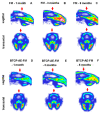
Electrospinning has recently been recognized as a potential method for use in biomedical applications such as nanofiber-based drug delivery or tissue engineering scaffolds. The present study aimed to demonstrate the electrospinning preparation and suitability of β-tricalcium phosphate-modified aerogel containing polyvinyl alcohol/chitosan fibrous meshes (BTCP-AE-FMs) for bone regeneration under in vitro and in vivo conditions. The mesh physicochemical properties included a 147 ± 50 nm fibrous structure, in aqueous media the contact angles were 64.1 ± 1.7°, and it released Ca, P, and Si. The viability of dental pulp stem cells on the BTCP-AE-FM was proven by an alamarBlue assay and with a scanning electron microscope. Critical-size calvarial defects in rats were performed as in vivo experiments to investigate the influence of meshes on bone regeneration. PET imaging using 18F-sodium fluoride standardized uptake values (SUVs) detected 7.40 ± 1.03 using polyvinyl alcohol/chitosan fibrous meshes (FMs) while 10.72 ± 1.11 with BTCP-AE-FMs after 6 months. New bone formations were confirmed by histological analysis. Despite a slight change in the morphology of the mesh because of cross-linking, the BTCP-AE-FM basically retained its fibrous, porous structure and hydrophilic and biocompatible character. Our experiments proved that hybrid nanospun scaffold composite mesh could be a new experimental bone substitute bioactive material in future medical practice.

Keywords: aerogel; electrospinning; electrospun meshes; tissue engineering; β-tricalcium phosphate.


Conflict of interest statement

The authors declare that they have no known competing financial interest or personal relationships that could have appeared to influence the work reported in this paper.

Figures
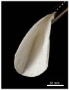
Figure 1
Figure 1
An electrospun BTCP-AE-FM folded in half.
Figure 2
Figure 2
Characterization of the different types of meshes: (a) representative SEM micrographs of a non-cross-linked FM, (b) a cross-linked FM, and (c) a cross-linked BTCP-AE-FM. Their diameter frequency distributions are shown on (df) bar graphs, respectively.
Figure 3
Figure 3
SEM microphotographs of the BTCP-AE powder sample (a) and BTCP-AE-FM sample (c). EDS analysis of the BTCP-AE inorganic composite powder sample (b) and BTCP-AE particle in the composite meshes (d).
Figure 4
Figure 4
Water contact angles of the BTCP-AE-FMs before and after cross-linking.
Figure 5
Figure 5
(a) IR spectra of the BTCP-AE-FM and FM fibrous meshes. The blue arrow indicates the pronounced presence of amide bonds in the aerogel composite mesh. (b) IR spectra of the starting materials showing highly overlapping peaks in the fingerprint region.
Figure 6
Figure 6
Recorded and best-fit ATR-FTIR spectra of the BTCP-AE-FM nanofiber composite assuming that the spectrum is the linear combination of the spectra of the FM and BTCP-AE. The blue arrow indicates the absorbance drop of the ester group ν(C-O) stretching frequencies.
Figure 7
Figure 7
(a) The leachable part of the BTCP-AE-FM in water. (b) The total amount of Ca, P, and Si ions in water after soaking periods. (c) The molar ratio of dissolved Ca and P. The time axis is shown in log scale in plots (a,c).
Figure 8
Figure 8
Cell viability assay of DPSC cells: cells were cultured with (CM+ and OM+) or without (CM and OM) BTCP-AE-FM samples for 7 and 14 days. After the incubation period, cell viability was assessed by alamarBlue assay. Values are expressed as sample means; error bars represent the standard deviation (SD) of three parallel measurements. In the t-tests, ** denotes p < 0.01, and **** denotes p < 0.0001. CM: control medium. OM: osteoinductive medium.
Figure 9
Figure 9
Live–dead assay of DPSCs. Cells were seeded onto 12-well plates and cultured with (CM+ and OM+) or without (CM and OM) BTCP-AE-FM samples for 7 and 14 days. After the incubation period, cells on the different surfaces were co-stained with fluorescein diacetate and propidium iodide.
Figure 10
Figure 10
SEM micrographs of DPSCs on the surface of the BTCP-AE-FM (a,b).
Figure 11
Figure 11
3D surface-rendered micro-CT reconstructions (axial view) with blue-colored ROI mask (8 mm). (AC) The FM and (DF) BTCP-AE-FM 1–3–6 months.
Figure 12
Figure 12
In vivo PET imaging of rat calvaria using [18F]fluoride. Representative decay-corrected PET images were obtained 1, 3, and 6 months after surgery and 50 min following intravenous injection of the radiopharmaceutical. (AC): sagittal and transaxial PET images of rats implanted with the FM; (DF): sagittal and transaxial PET images of rats implanted with the BTCP-AE-FM. Red arrows: area of the cranial trepanation surgery.
Figure 13
Figure 13
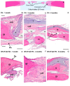
Histological analysis of bone remodeling in HE-stained rat calvaria plates. (A) The schematic diagram explains multifocal bone remodeling during calvarial defect repair. Progress of reossification was demonstrated with HE staining in the critical defect, applying polyvinyl alcohol and chitosan blended fibrous mesh (FM) (BD) and inorganic composite modified polyvinyl alcohol and chitosan hybrid fibrous mesh (BTCP-AE-FM) (EG) implants. Magnification: 10× obj (B,C,E,F); 4× obj (D,G). Pe: periosteum. Of: osteogenic front. is: bone islets. Coll: collagenous tissue. Ves: blood vessels. *: implanted fibrous mesh. Scalebar: 0.5 mm (B,C,E,F); 1 mm (D,G).
Figure 14
Figure 14
Residual silica compounds following mesoporous BTCP-AE insertion into the calvaria bone defect 6 months after treatment. The hematoxylin-eosin (HE) stained image (left) shows sub-total re-ossification of the defect at this time, highlighted by the solid hypocellular pink tissue. However, the center of the picture still mainly exhibits an inflammatory fibrous non-ossified region with the presence of a foreign body giant cell granulomatous reaction in association with the presence of non-metabolized silica crystal particle remnants (within the black frame). This region is magnified (630×) in the right-hand side image, which was photographed in a polarizing microscope to confirm the presence of crystalloid foreign particles (the birefringent bright white materials, indicated by the blue arrows) surrounded by the epithelioid-activated macrophages (thin red arrows) and the multinuclear (foreign body) giant cells (thick red arrows) [32].
Figure 15
Figure 15
Workflow of the fabrication, characterization, and in vitro and in vivo investigations of chemically cross-linked biodegradable BTCP-AE-FMs.
Figure 16
Figure 16
Contact angle measurement of a water droplet (2 µL) on a BTCP-AE-FM sample.
Figure 17
Figure 17
The surgical process for the critical size calvarial defects in rats. (a) Preparation of the 8 mm diameter full-thickness defect. (b) Surgical area after bone removal. (c) Implanted BTCP-AE-FM plate in the defective site.

References

    1. Putra R.U., Basri H., Prakoso A.T., Chandra H., Ammarullah M.I., Akbar I., Syahrom A., Kamarul T. Level of Activity Changes Increases the Fatigue Life of the Porous Magnesium Scaffold, as Observed in Dynamic Immersion Tests, over Time. Sustainability. 2023;15:823. doi: 10.3390/su15010823. - DOI
    1. Qi Y., Wang C., Wang Q., Zhou F., Li T., Wang B., Su W., Shang D., Wu S. A Simple, Quick, and Cost-Effective Strategy to Fabricate Polycaprolactone/Silk Fibroin Nanofiber Yarns for Biotextile-Based Tissue Scaffold Application. Eur. Polym. J. 2023;186:111863. doi: 10.1016/j.eurpolymj.2023.111863. - DOI
    1. Nazarnezhad S., Baino F., Kim H.W., Webster T.J., Kargozar S. Electrospun Nanofibers for Improved Angiogenesis: Promises for Tissue Engineering Applications. Nanomaterials. 2020;10:1609. doi: 10.3390/nano10081609. - DOI - PMC - PubMed
    1. Li M., Qiu W., Wang Q., Li N., Liu L., Wang X., Yu J., Li X., Li F., Wu D. Nitric Oxide-Releasing Tryptophan-Based Poly(Ester Urea)s Electrospun Composite Nanofiber Mats with Antibacterial and Antibiofilm Activities for Infected Wound Healing. ACS Appl. Mater. Interfaces. 2022;14:15911–15926. doi: 10.1021/acsami.1c24131. - DOI - PubMed
    1. Prakoso A.T., Basri H., Adanta D., Yani I., Ammarullah M.I., Akbar I., Ghazali F.A., Syahrom A., Kamarul T. The Effect of Tortuosity on Permeability of Porous Scaffold. Biomedicines. 2023;11:427. doi: 10.3390/biomedicines11020427. - DOI - PMC - PubMed

LinkOut - more resources