. 2023 Apr 20;24(8):7589.
doi: 10.3390/ijms24087589.

Mitoregulin Contributes to Creatine Shuttling and Cardiolipin Protection in Mice Muscle

Affiliations

Mitoregulin Contributes to Creatine Shuttling and Cardiolipin Protection in Mice Muscle

Olga A Averina et al. Int J Mol Sci. .

Abstract

Small peptides compose a large share of the mitochondrial proteome. Mitoregulin (Mtln) is a mitochondrial peptide known to contribute to the respiratory complex I functioning and other processes in mitochondria. In our previous studies, we demonstrated that Mtln knockout mice develop obesity and accumulate triglycerides and other oxidation substrates in serum, concomitant with an exhaustion of tricarboxylic acids cycle intermediates. Here we examined the functional role of Mtln in skeletal muscles, one of the major energy consuming tissues. We observed reduced muscle strength for Mtln knockout mice. Decrease of the mitochondrial cardiolipin and concomitant increase in monolysocardiolipin concentration upon Mtln inactivation is likely to be a consequence of imbalance between oxidative damage and remodeling of cardiolipin. It is accompanied by the mitochondrial creatine kinase octamer dissociation and suboptimal respiratory chain performance in Mtln knockout mice.

Keywords: cardiolipin; creatine kinase; metabolism; mitochondria; oxidative phosphorylation; short open reading frame; small peptide.


Conflict of interest statement

The authors declare no conflict of interest.

Figures

Figure 1
Figure 1
Influence of Mtln inactivation on integrative characteristics of muscle. (a) Forelimb grip strength of the wild type (green bar, n = 10) and ΔMtln-1 (red bar, n = 13) male mice after 24 h food deprivation; interquartile ranges are shown as solid bars, while the all-data range is shown by thin lines. The horizontal line corresponds to the median, while the cross to the average. The significance level calculated using the Student’s t-test is shown; If a p-value is less than 0.05, it is flagged with one star (*) (b) Differential gene expression in the soleus muscle of the wild type and ΔMtln-1 knockout mice. Volcano plot of differentially expressed genes. The x-axis corresponds to the log-scale fold change of expression, ΔMtln-1 relative to the wild type, while the y-axis corresponds to p-value.
Figure 2
Figure 2
Influence of Mtln gene inactivation on respiration of muscle mitochondria. Oxygen consumption rate (OCR) of soleus muscle mitochondria extracted from the wild type (green bars, n = 3) and ΔMtln-1 (red bars, n = 3) male mice. The groups of bars correspond to the respiration on palmitoyl carnitine (CI + CII + ETF activity), pyruvate and malate (CI activity), glutamate and malate (CI activity) and succinate (CII activity) as marked below the graphs. The experimental points measured are substrates alone (init), substrates with rotenone (rot), substrates and ADP (ADP (3)), substrates after exhaustion of ADP (ADP (4)), substrates after exhaustion of ADP after addition of oligomycin (ADP (4o)), substrates after exhaustion of ADP (ADP (4)) following addition of creatine, substrates after exhaustion of ADP after addition of oligomycin and FCCP uncoupler (ADP (3u) FCCP), residual respiration after inhibition of uncoupled respiration by potassium cyanide. Interquartile ranges are shown as solid bars, while the all-data range is shown by thin lines. The horizontal line corresponds to the median, while the cross is the average. The significance level calculated using the Student’s t-test is shown. If a p-value is less than 0.05, it is flagged with one star (*).
Figure 3
Figure 3
Influence of Mtln gene inactivation on creatine kinase functioning. (a) Relative abundance of the octameric and dimeric forms of the mitochondrial creatine kinase (mtCK) in the soleus muscle mitochondrial extracts of the wild type (green bars, n = 18) and ΔMtln-1 (red bars, n = 10) male mice; (b) Mitochondrial (left group of bars) and cytosolic (right group of bars) creatine kinase activity for the soleus extracts of the wild type (green bars, n = 18) and ΔMtln-1 (red bars, n = 10) male mice. For all panels, interquartile ranges are shown as solid bars, while the all-data range is shown by thin lines. The horizontal line corresponds to the median, while the cross is the average. Significance level calculated using the Student’s t-test is shown. If a p-value is less than 0.01, it is flagged with two stars (**), while four stars (****) corresponds to a p-value less than 10−4.
Figure 4
Figure 4
Influence of Mtln gene inactivation on cardiolipin amount and integrity. (a) Cardiolipin quantitation in the soleus mitochondria of the wild type (green bars, n = 6) and ΔMtln-1 (red bars, n = 7) mice; (b) Quantitation of the amount of monolysocardiolipin (MLCL) relative to the total amount of cardiolipin and monolysocardiolipin (MLCL+CL) in the soleus mitochondria of the wild type (green bars, n = 7) and ΔMtln-1 (red bars, n = 8) mice; (c) Thin layer chromatography of the soleus mitochondrial lipids of the wild type male (left 3 lanes) and ΔMtln-1 male (right 4 lanes) mice. Lipid designations are: fatty acids (FA), cardiolipin (CL), monolysocardiolipin (MLCL), phosphatidic acid (PA), phosphatidylethanolamine (PE), phosphatidylinositol (PI), phosphatidylserine (PS), phosphatidylcholine (PC), lysophosphatidylcholine (LPC). For panels a and b, interquartile ranges are shown as solid bars, while the all-data range is shown by thin lines. The horizontal line corresponds to the median, while the cross is the average. Significance level calculated using the Student’s t-test is shown. If a p-value is less than 10−4, it is flagged with four stars.
Figure 5
Figure 5
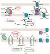
A model for the Mtln function. (a) Mitochondrial homeostasis in the presence of intact Mtln. Shown is an interaction of Mtln (pink) with cardiolipin (CL) and CYB5R3 NADH dehydrogenase preventing oxidative lipid damage by the reactive oxygen species (ROS). It ensures normal CL concentration and proper activity of the respiratory complex I (I). The tricarboxylic acids (TCA) cycle functions normally and ensures oxidation of substrates. Cardiolipin facilitates mitochondrial creatine kinase (mtCK) octamerization in the intermembrane space coupling ATP/ADP antiporter (ANT) activity, creatine (Cr) phosphorylation and creatine phosphate (CrP) export to the cytosol via VDAC. (b) Consequences of Mtln inactivation. Lack of Mtln leads to a loss of its interaction with CL and increased ROS production, likely due to the decreased CYB5R3 activity. Increased concentrations of ROS cause excessive CL damage. Monolysocardiolipin (MLCL), an intermediate of CL repair is accumulating. Decrease in the functional CL concentration leads to suboptimal function of CI which leads to the accumulation of substrates of respiration and depletion of its products and TCA cycle intermediates. Red downward arrows mark metabolites whose concentration is reduced or enzymes whose activity is reduced upon Mtln inactivation, while upward green arrows mark those metabolites whose concentration was increased. The CL concentration drop affects mtCK octamer formation and consequently the Cr/CrP dependent energy relay efficiency.

Similar articles

Cited by

References

    1. Chugunova A., Navalayeu T., Dontsova O., Sergiev P. Mining for Small Translated ORFs. J. Proteome Res. 2018;17:1–11. doi: 10.1021/acs.jproteome.7b00707. - DOI - PubMed
    1. Sergiev P.V., Rubtsova M.P. Little but Loud. The Diversity of Functions of Small Proteins and Peptides—Translational Products of Short Reading Frames. Biochem. Mosc. 2021;86:1139–1150. doi: 10.1134/S0006297921090091. - DOI - PubMed
    1. Couso J.-P., Patraquim P. Classification and Function of Small Open Reading Frames. Nat. Rev. Mol. Cell Biol. 2017;18:575–589. doi: 10.1038/nrm.2017.58. - DOI - PubMed
    1. Makarewich C.A., Olson E.N. Mining for Micropeptides. Trends Cell Biol. 2017;27:685–696. doi: 10.1016/j.tcb.2017.04.006. - DOI - PMC - PubMed
    1. Ruiz-Orera J., Albà M.M. Translation of Small Open Reading Frames: Roles in Regulation and Evolutionary Innovation. Trends Genet. 2019;35:186–198. doi: 10.1016/j.tig.2018.12.003. - DOI - PubMed

LinkOut - more resources