. 2023 Apr 21;24(8):7627.
doi: 10.3390/ijms24087627.

Can Neutrophils Prevent Nosocomial Pneumonia after Serious Injury?

Affiliations

Can Neutrophils Prevent Nosocomial Pneumonia after Serious Injury?

Kristína Macáková et al. Int J Mol Sci. .

Abstract

Nosocomial pneumonia is a leading cause of critical illness and mortality among seriously injured trauma patients. However, the link between injury and the development of nosocomial pneumonia is still not well recognized. Our work strongly suggests that mitochondrial damage-associated molecular patterns (mtDAMPs), especially mitochondrial formyl peptides (mtFPs) released by tissue injury, play a significant role in developing nosocomial pneumonia after a serious injury. Polymorphonuclear leukocytes (neutrophils, PMN) migrate toward the injury site by detecting mtFPs through formyl peptide receptor 1 (FPR1) to fight/contain bacterial infection and clean up debris. Activation of FPR1 by mtFPs enables PMN to reach the injury site; however, at the same time it leads to homo- and heterologous desensitization/internalization of chemokine receptors. Thus, PMN are not responsive to secondary infections, including those from bacteria-infected lungs. This may enable a progression of bacterial growth in the lungs and nosocomial pneumonia. We propose that the intratracheal application of exogenously isolated PMN may prevent pneumonia coupled with a serious injury.

Keywords: FPR1; infection; injury; innate immunity; neutrophils; nosocomial pneumonia; trauma.


Conflict of interest statement

The authors declare no conflict of interest.

Figures

Figure 1
Figure 1
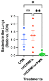
Effects of mtDAMPs and external mouse PMN on S. aureus (SA) clearance in lung. Left: CD-1 mice were separated into three groups. 1. CON: Saline i.p. injection (time 0) followed by S. aureus (3 h) i.t. 2. mtDAMPs: mtDAMPs i.p. followed by S. aureus, 3. mtDAMPs + PMN: mtDAMPs i.p. followed by S. aureus and BM-PMN i.t. (6 h). Animals were sacrificed t = 24 h. Lung homogenates were prepared to determine bacterial presence. mtDAMPs prepared from 10% of total liver in saline. 50 µL of OD600 = 0.1 S. aureus was injected intratracheally. BM-PMN were freshly prepared from donor CD-1 mice and ~2 × 10⁶ cells were injected intratracheally. The numbers of animals used for CON, mtDAMPs, and mtDAMPs + PMN were N = 8, N = 8, and N = 9, respectively. Mean and SE values are shown. * denotes a significant difference by one-way ANOVA, Tukey [14]. *: p = 0.027, **: p = 0.015. ns: not significant. Right: The animal protocol as early onset with S. aureus.
Figure 2
Figure 2
Effects of mtDAMPs and external mouse PMN on P. aeruginosa clearance (PA) in lung. Left: CD-1 mice were separated into three groups. 1. CON: Saline i.p. injection (time 0) followed by P. aeruginosa (24 h) i.t. 2. mtDAMPs: mtDAMPs i.p. (time 0) followed by P. aeruginosa i.t. (24 h), 3. mtDAMPs + PMN: mtDAMPs i.p. (time 0) followed by P. aeruginosa i.t. (24 h) and BM-PMN i.t. (27 h). Animals were sacrificed t = 48 h. Lung homogenates were prepared to determine bacterial presence. mtDAMPs were prepared from 10% of total liver in saline. A total of 50 μL of OD600 = 0.1, P. aeruginosa was injected intratracheally. BM-PMN were freshly prepared from donor CD-1 mice and ~2 × 10⁶ cells were injected intratracheally. The numbers of animals used for CON, mtDAMPs, and mtDAMPs + PMN were N = 10, N = 11, and N = 12, respectively. Mean and SE values are shown. * denotes a significant difference by one-way ANOVA, Tukey [14]. *: p = 0.047, **: p = 0.038. ns: not significant. Right: The animal protocol as late onset with P. aeruginosa.
Figure 3
Figure 3
Effects of exogenous PMN on clearance of Pseudomonas pneumonia. Protocols are similar to Figure 2; however, an increased number of P. aeruginosa (OD = 0.111) was injected to the lungs. The numbers of animals used for CON, mtDAMPs, and mtDAMPs + PMN were N = 4, N = 6, and N = 8, respectively. Mean and SE values are shown. *: denotes a significant difference by one-way ANOVA, Tukey [14]. *: p = 0.036, **: p = 0.004. ns: not significant.
Figure 4
Figure 4
Exogenous PMN do not injure the lung. Lung injury was evaluated by assaying protein leak into BAL fluids. CON: saline i.t. (n = 5), CD-1 to CD-1: BM-PMN from CD-1 mice were instilled i.t. into CD-1 mice, 2 × 10⁶ i.t. (n = 5), CD-1 to BL6: BM-PMN from CD-1 mice were instilled i.t. into BL6 mice (n = 2). BL6 to CD-1: BM-PMN from BL6 mice (n = 2) were instilled i.t. into CD-1 mice. mtDAMPs/Bac/HBSS: mtDAMPs from 10% liver is given i.p. at t = 0. S. aureus is given i.t. (8.6 × 10⁶ CFU) at T = 3 h and followed by HBSS (vehicle for PMN) (n = 3). mtDAMPs/Bac/PMN: As in mtDAMPs/Bac but followed by PMN i.t. (1 × 10⁶) at T = 6 h (n = 3). *: denotes a significant difference by one-way ANOVA with Tukey’s test. ns denotes p = 0.090. ****: p < 0.0001. ns: not significant. Sample collection: Control (saline) and CD-1 to CD-1 PMN i.t. 24 h, BL6 to CD-1 or CD-1 to BL6: 72 h, mtDAMPs/Bac/HBSS, and mtDAMPs/Bac/PMN: 23 h. The number in bracket represents the number of animals used [14].
Figure 5
Figure 5
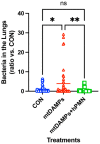
Effects of mtDAMPs and external human PMN on S. aureus (SA) clearance in lung. C57BL6 mice were separated into three groups. Protocols are similar to Figure 1. 1. CON: Saline i.p. injection (time 0) followed by S. aureus (3 h) i.t. 2. mtDAMPs: mtDAMPs i.p. (time 0) followed by S. aureus i.t. (3 h) and saline i.t. (6 h). 3. mtDAMPs + hPMN: mtDAMPs i.p. (time 0) followed by S. aureus i.t. (3 h) and human PMN i.t. (6 h). Animals were sacrificed t = 20 h. Lung homogenates were prepared to determine bacterial presence. MTD were prepared from 10% of total liver in saline. A total of 50 μL of OD600 = 0.1, S. aureus was applied intratracheally. Human PMN were freshly prepared from healthy donor and ~2 × 10⁶ cells were injected intratracheally. The numbers of animals used for CON, mtDAMPs, and mtDAMPs + PMN were n = 18, n = 21, and n = 21, respectively. Mean and SE values are shown. *: denotes a significant difference by one-way ANOVA, Tukey [44]. *: p = 0.0129, **: p = 0.0020. ns: not significant.
Figure 6
Figure 6
Reactive oxygen species production by freeze–thaw PMN. Freeze–thaw human PMN were loaded with Luminor and 100 nM fMLF (red), PMA (green), or buffer (blue) were applied to stimulate PMN to produce reactive oxygen species (ROS). Real time ROS production is shown in (A). Area under curve (AUC) for 5200 s was calculated to compare the ROS production (B). Experiments were done in quadruplicates. Mean and SE values are shown. *: denotes a significant difference by one-way ANOVA, Tukey. ****: p < 0.0001.
Figure 7
Figure 7
Freeze–thaw PMN induce bacterial killing in vivo. Similar to Figure 5, we applied freshly isolated and freeze–thaw human PMN to our mouse injury/lung bacterial infection model. Although there was no significant difference (ns), we could see a tendency that freeze–thaw PMN could be as effective as freshly isolated human PMN. The numbers of animals used for control (CTRL), mtDAMPs, mtDAMPs + fresh PMN (Fresh PMN), and mtDAMPs + freeze–thaw PMN (F-T PMN) were n = 4, n = 7, and n = 8, and n = 8, respectively. Mean and SE values are shown. There was no significant difference by one-way ANOVA, Tukey.

Similar articles

Cited by

References

    1. Chevret S., Hemmer M., Carlet J., Langer M. Incidence and risk factors of pneumonia acquired in intensive care units. Results from a multicenter prospective study on 996 patients. European Cooperative Group on Nosocomial Pneumonia. Intensive Care Med. 1993;19:256–264. - PubMed
    1. Papia G., Louie M., Tralla A., Johnson C., Collins V., Simor A.E. Screening high-risk patients for methicillin-resistant Staphylococcus aureus on admission to the hospital: Is it cost effective? Infect. Control Hosp. Epidemiol. 1999;20:473–477. doi: 10.1086/501655. - DOI - PubMed
    1. Papia G., McLellan B.A., El-Helou P., Louie M., Rachlis A., Szalai J.P., Simor A.E. Infection in hospitalized trauma patients: Incidence, risk factors, and complications. J. Trauma. 1999;47:923–927. doi: 10.1097/00005373-199911000-00018. - DOI - PubMed
    1. Wallace W.C., Cinat M., Gornick W.B., Lekawa M.E., Wilson S.E. Nosocomial infections in the surgical intensive care unit: A difference between trauma and surgical patients. Am. Surg. 1999;65:987–990. doi: 10.1177/000313489906501019. - DOI - PubMed
    1. Kozka M., Sega A., Wojnar-Gruszka K., Tarnawska A., Gniadek A. Risk Factors of Pneumonia Associated with Mechanical Ventilation. Int. J. Environ. Res. Public Health. 2020;17:656. doi: 10.3390/ijerph17020656. - DOI - PMC - PubMed