. 2023 Apr 21;24(8):7660.
doi: 10.3390/ijms24087660.

Inflammatory Response and Exosome Biogenesis of Choroid Plexus Organoids Derived from Human Pluripotent Stem Cells

Affiliations

Inflammatory Response and Exosome Biogenesis of Choroid Plexus Organoids Derived from Human Pluripotent Stem Cells

Laureana Muok et al. Int J Mol Sci. .

Abstract

The choroid plexus (ChP) is a complex structure in the human brain that is responsible for the secretion of cerebrospinal fluid (CSF) and forming the blood-CSF barrier (B-CSF-B). Human-induced pluripotent stem cells (hiPSCs) have shown promising results in the formation of brain organoids in vitro; however, very few studies to date have generated ChP organoids. In particular, no study has assessed the inflammatory response and the extracellular vesicle (EV) biogenesis of hiPSC-derived ChP organoids. In this study, the impacts of Wnt signaling on the inflammatory response and EV biogenesis of ChP organoids derived from hiPSCs was investigated. During days 10-15, bone morphogenetic protein 4 was added along with (+/-) CHIR99021 (CHIR, a small molecule GSK-3β inhibitor that acts as a Wnt agonist). At day 30, the ChP organoids were characterized by immunocytochemistry and flow cytometry for TTR (~72%) and CLIC6 (~20%) expression. Compared to the -CHIR group, the +CHIR group showed an upregulation of 6 out of 10 tested ChP genes, including CLIC6 (2-fold), PLEC (4-fold), PLTP (2-4-fold), DCN (~7-fold), DLK1 (2-4-fold), and AQP1 (1.4-fold), and a downregulation of TTR (0.1-fold), IGFBP7 (0.8-fold), MSX1 (0.4-fold), and LUM (0.2-0.4-fold). When exposed to amyloid beta 42 oligomers, the +CHIR group had a more sensitive response as evidenced by the upregulation of inflammation-related genes such as TNFα, IL-6, and MMP2/9 when compared to the -CHIR group. Developmentally, the EV biogenesis markers of ChP organoids showed an increase over time from day 19 to day 38. This study is significant in that it provides a model of the human B-CSF-B and ChP tissue for the purpose of drug screening and designing drug delivery systems to treat neurological disorders such as Alzheimer's disease and ischemic stroke.

Keywords: Wnt signaling; choroid plexus organoids; extracellular vesicles; human pluripotent stem cells; inflammatory response.


Conflict of interest statement

No competing financial interest exist.

Figures

Figure 1
Figure 1
ChP organoid differentiation from hiPSCs. (A) Illustration of ChP differentiation; (B) the ChP organoid morphology for CHIR+/− conditions during the differentiation. Scale bar: 200 µm. BMP: bone morphogenetic protein; EBs: embryoid bodies. EV: extracellular vesicles.
Figure 2
Figure 2
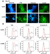
Expression of ChP markers for CHIR+/− conditions. (A) Immunostaining of ChP markers at day 32. Scale bar: 50 µm. (B) Flow cytometry histograms of ChP markers for CHIR+/− conditions. The assays were performed at day 30. Black line: negative control. Red line: marker of interest. TTR (transthyretin) and CLIC6 (chloride intracellular channel 6) are ChP markers. CHIR(+/−) indicates treatment with CHIR99021.
Figure 3
Figure 3
mRNA expression of ChP markers at days 18 and 30 for CHIR+/− conditions. mRNA expression was determined by RT-qPCR. (A) Day 18; (B) day 30 of differentiation. N = 3. * indicates p < 0.05. CHIR(+/−) indicates treatment with CHIR99021.
Figure 4
Figure 4
Inflammatory response to Aβ42 oligomer stimulation for ChP organoids of CHIR+/− conditions. mRNA expression determined by RT-qPCR for (A) proinflammatory markers (day 33); (B) anti-inflammatory markers (day 33); these markers were relative to the CHIR+ condition. (C) MMP2, MMP3, and MMP9 expression affected by Aβ42 oligomers (day 33); (D) MMP2, MMP3, and MMP9 expression affected by CHIR at days 19 and 38. N = 3. * indicates p < 0.05. The MMP2, MMP3, and MMP9 expression was relative to the CHIR−D19 condition. CHIR(+/−) indicates treatment with CHIR99021, D19 and D38 indicates days 19 and 38, AB(+/−) indicates treatment with amyloid β 42 oligomers. The CHIR+ group is more responsive than the CHIR− group.
Figure 5
Figure 5
Extracellular vesicle biogenesis of ChP organoids with CHIR+/− conditions. (A) mRNA expression determined by RT-qPCR for (i) ESCRT-dependent EV biogenesis markers; (ii) ESCRT-independent EV biogenesis markers; N = 3. * indicates p < 0.05 for the compared conditions, and # indicates p < 0.05 compared to the CHIR− condition at the same time point. CHIR(+/−) indicates treatment with CHIR99021 (3 µM), D19 and D38 indicates days 19 and 38 during differentiation. (B) Nanoparticle tracking analysis (NTA) for the isolated ChP organoid-EVs; (i) particle size distribution and zeta potential by ZetaView; (ii) particle size distribution by NanoSight. (C) Transmission electron microscopy images showing exosome morphology; scale bar: 100 nm.

References

    1. Lun M.P., Monuki E.S., Lehtinen M.K. Development and functions of the choroid plexus-cerebrospinal fluid system. Nat. Rev. Neurosci. 2015;16:445–457. doi: 10.1038/nrn3921. - DOI - PMC - PubMed
    1. Lehtinen M.K., Bjornsson C.S., Dymecki S.M., Gilbertson R.J., Holtzman D.M., Monuki E.S. The choroid plexus and cerebrospinal fluid: Emerging roles in development, disease, and therapy. J. Neurosci. 2013;33:17553–17559. doi: 10.1523/JNEUROSCI.3258-13.2013. - DOI - PMC - PubMed
    1. Jacob F., Pather S.R., Huang W.K., Zhang F., Wong S.Z.H., Zhou H., Cubitt B., Fan W., Chen C.Z., Xu M., et al. Human Pluripotent Stem Cell-Derived Neural Cells and Brain Organoids Reveal SARS-CoV-2 Neurotropism Predominates in Choroid Plexus Epithelium. Cell Stem Cell. 2020;27:937–950.e939. doi: 10.1016/j.stem.2020.09.016. - DOI - PMC - PubMed
    1. Zhao J., Fu Y., Yamazaki Y., Ren Y., Davis M.D., Liu C.C., Lu W., Wang X., Chen K., Cherukuri Y., et al. APOE4 exacerbates synapse loss and neurodegeneration in Alzheimer’s disease patient iPSC-derived cerebral organoids. Nat. Commun. 2020;11:5540. doi: 10.1038/s41467-020-19264-0. - DOI - PMC - PubMed
    1. Garreta E., Kamm R.D., Chuva de Sousa Lopes S.M., Lancaster M.A., Weiss R., Trepat X., Hyun I., Montserrat N. Rethinking organoid technology through bioengineering. Nat. Mater. 2021;20:145–155. doi: 10.1038/s41563-020-00804-4. - DOI - PubMed

Substances